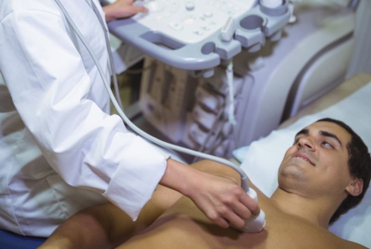
The USG Chest Scan, an ultrasound examination tailored for thoracic imaging, offers a non-invasive approach to explore the complexities of the chest cavity.
Ultrasound / USG Chest Scan in India with Cost
Ultrasound or USG Chest
Scan in Detail: Navigating the Intricacies of Thoracic Imaging
Exploring the realm of diagnostic imaging, the Ultrasound or USG (Ultrasonography) Chest Scan emerges as a valuable tool for examining the thoracic region. This comprehensive guide delves into the significance, procedure, and insights provided by the USG Chest Scan, shedding light on its role in evaluating thoracic structures.
Introduction
The USG Chest Scan, an ultrasound examination tailored for thoracic imaging, offers a non-invasive approach to explore the complexities of the chest cavity. This article aims to unravel the intricacies of the procedure, emphasizing its importance in diagnosing and monitoring various thoracic conditions.
Understanding USG Chest Scan
Unlike traditional chest imaging methods such as X-rays or CT scans, the USG Chest Scan employs sound waves to create real-time images of the thoracic structures. It serves as a valuable diagnostic tool for assessing organs like the lungs, heart, and surrounding tissues.
Importance in Diagnostic Imaging
The USG Chest Scan plays a crucial role in thoracic diagnostic imaging, allowing healthcare providers to evaluate and diagnose a range of conditions affecting the chest. Its non-invasive nature and real-time imaging capabilities contribute to its significance in medical diagnostics.
Preparation for the Scan
Preparation for a USG Chest Scan is typically minimal. Patients may be instructed to wear comfortable clothing and may need to remove any jewelry or accessories that could interfere with the ultrasound probe. The procedure is generally well-tolerated, requiring no special dietary restrictions.
Procedure: A Window into
Thoracic Structures
Gel Application: A clear gel is applied to the chest area,
facilitating the transmission of sound waves.
Transducer Movements: The transducer,
a handheld device, is gently moved over the chest, emitting sound waves that
bounce back to create detailed images.
Real-time Imaging: The procedure
provides real-time images of thoracic structures, allowing healthcare providers
to assess movement and function.
Assessment Areas in
Thoracic Imaging
The USG Chest Scan is adept at assessing various thoracic structures, including:
Lungs: Examining lung tissue for
abnormalities or fluid accumulation.
Heart: Assessing the heart's structure
and function.
Pleura: Evaluating the pleural space for
signs of inflammation or effusion.
Blood Vessels: Examining blood vessels for
abnormalities or blockages.
Thoracic Wall: Assessing the integrity of the
chest wall and surrounding tissues.
Benefits of USG Chest
Scan
Non-Invasiveness: The procedure is non-invasive, avoiding exposure to ionizing radiation.
Real-time Imaging: Provides dynamic, real-time images for immediate assessment.
Focused Evaluation: Allows for focused evaluation of specific thoracic structures.
Risks and Considerations
USG Chest Scan is considered a safe procedure with minimal risks. It is particularly advantageous for certain patient populations, such as pregnant individuals or those sensitive to ionizing radiation.
Clinical Applications
The USG Chest Scan finds utility in diagnosing and monitoring various thoracic conditions, including:
Pleural Effusion: Detecting abnormal fluid accumulation in the
pleural space.
Pneumothorax: Identifying air leaks in the
pleural cavity.
Cardiac Conditions: Assessing the
heart's structure and function.
Pulmonary Pathologies: Evaluating lung
tissue for abnormalities.
Expert Perspectives
Medical professionals emphasize the role of the USG Chest Scan in providing valuable diagnostic information without the use of ionizing radiation. Their insights contribute to informed decision-making and patient care.
Technological Advancements
Advancements in ultrasound technology continually enhance the precision and clarity of the USG Chest Scan, providing more detailed and nuanced information for diagnostic purposes.
Patient Experience
The USG Chest Scan is generally well-tolerated by patients, offering a comfortable and efficient imaging experience. Its non-invasive nature contributes to a positive patient experience.
Conclusion
In conclusion, the Ultrasound or USG Chest Scan stands as a valuable diagnostic tool for thoracic imaging, providing real-time insights into the complexities of the chest cavity. Its non-invasive nature, real-time imaging capabilities, and focus on specific thoracic structures contribute to its importance in diagnostic imaging.
FAQs (Frequently Asked Questions) related to the Ultrasound or USG Chest
Scan
1. Can a USG Chest Scan detect lung cancer?
While the USG Chest Scan is useful for assessing lung tissues, it may not be the primary tool for detecting lung cancer. Additional imaging or diagnostic tests may be recommended based on clinical suspicion.
2. Is the USG Chest Scan safe during pregnancy?
Yes, the USG Chest Scan is considered safe during pregnancy due to its non-invasive nature and lack of ionizing radiation. However, specific considerations may be discussed with the healthcare provider.
3. How long does a USG Chest Scan typically take?
The duration of a USG Chest Scan varies but is generally a relatively quick procedure, typically taking around 15 to 30 minutes.
4. Can the USG Chest Scan identify blood clots in the pulmonary arteries?
While the USG Chest Scan is adept at assessing blood vessels, it may not be the primary method for diagnosing blood clots in the pulmonary arteries. Complementary imaging studies such as CT pulmonary angiography are often employed for this purpose.
5. Is there any special preparation required before a USG Chest Scan?
Typically, there is minimal preparation required for a USG Chest Scan. Patients may be advised to wear comfortable clothing and, in some cases, refrain from applying lotions or creams to the chest area.
6. Can the USG Chest Scan be used to monitor the progress of respiratory conditions?
Yes, the USG Chest Scan can be instrumental in monitoring the progression of respiratory conditions, assessing lung function, and identifying any changes in thoracic structures.
7. Are there any age restrictions for undergoing a USG Chest Scan?
There are generally no age restrictions for a USG Chest Scan. This diagnostic tool is applicable to individuals of all ages, from infants to the elderly.
8. Can the USG Chest Scan detect abnormalities in the mediastinum?
Yes, the USG Chest Scan is capable of detecting abnormalities in the mediastinum, including tumors or masses in the central thoracic region.
9. Is the USG Chest Scan suitable for assessing heart valve function?
While the USG Chest Scan provides valuable information about the heart's structure, it may not be the primary method for assessing heart valve function. Echocardiography is often employed for detailed cardiac evaluations.
10. Does insurance typically cover the cost of a USG Chest Scan?
Coverage for a USG Chest Scan may vary based on individual insurance plans. It's advisable to check with the insurance provider to determine coverage and any associated costs.
These additional FAQs aim to provide more insights into the Ultrasound or USG Chest Scan, addressing common questions that individuals may have regarding the procedure.
(0)
Login to continue


