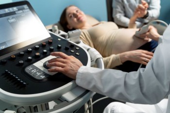
USG Single Joint imaging is a targeted ultrasound examination designed to assess the anatomy and pathology of a specific joint within the musculoskeletal system.
Ultrasound / USG Single Joint in India with Cost
USG Single Joint in
Detail: Peering into Precision for Musculoskeletal Insights
Within the realm of musculoskeletal diagnostics, the Ultrasound or USG (Ultrasonography) Single Joint examination emerges as a specialized technique, focusing on the detailed imaging of a specific joint. This comprehensive guide aims to illuminate the significance, procedure, and invaluable insights provided by USG Single Joint imaging in achieving precision in musculoskeletal diagnostics.
Introduction
USG Single Joint imaging is a targeted ultrasound examination designed to assess the anatomy and pathology of a specific joint within the musculoskeletal system. This technique allows for real-time visualization of joint structures, providing crucial information for diagnosing various conditions affecting the joints.
Understanding USG Single Joint Imaging
Unlike broader musculoskeletal ultrasound assessments, USG Single Joint imaging hones in on a particular joint, allowing for detailed examination of the joint's ligaments, tendons, synovial lining, and surrounding soft tissues. This targeted approach is instrumental in identifying joint-related issues and guiding appropriate medical interventions.
Importance in Musculoskeletal Diagnostics
USG Single Joint imaging holds paramount importance in the diagnosis and management of joint-related conditions. It offers a dynamic and detailed assessment, aiding healthcare providers in identifying structural abnormalities, inflammatory changes, and injuries within a specific joint.
Preparation for the Scan
Preparation for a USG Single Joint examination is generally straightforward. Patients may be advised to wear loose and comfortable clothing that allows easy access to the targeted joint. The procedure is typically well-tolerated, and no specific dietary restrictions or fasting are required.
Procedure: Precision in
Joint Imaging
Patient Positioning: Depending on the joint being examined, the
patient is positioned to optimize the visualization of the targeted area.
Gel Application: A water-based gel is applied to
the skin over the joint, facilitating the transmission of sound waves and
ensuring proper contact with the transducer.
Transducer Movements: The transducer,
a handheld device, is gently moved over the gel-coated skin, emitting and
receiving sound waves. This process creates real-time images of the joint's
internal structures.
Dynamic Evaluation: USG Single
Joint imaging allows for dynamic assessment, enabling the observation of joint
movement, the integrity of ligaments and tendons, and any abnormalities during
real-time imaging.
Assessment Areas in
Joint Imaging
USG Single Joint imaging is employed for the detailed evaluation of various joints, including but not limited to:
Knee Joint: Assessing ligaments, menisci, and
the synovial lining for conditions such as arthritis or tears.
Shoulder Joint: Examining tendons, the rotator
cuff, and bursae for issues like impingement or inflammation.
Elbow Joint: Evaluating structures such as the
ulnar collateral ligament or bursae for conditions like tennis elbow.
Ankle Joint: Assessing ligaments, tendons, and
joint spaces for issues such as sprains or arthritis.
Benefits of USG Single
Joint Imaging
Precision and Detail: Provides high-resolution
images, allowing for a detailed examination of specific joint structures.
Real-time Visualization: Enables dynamic
evaluation, capturing joint movements and abnormalities during motion.
Non-Invasive and Radiation-Free: USG imaging
does not involve ionizing radiation, ensuring patient safety.
Risks and Considerations
USG Single Joint imaging is generally considered a safe procedure with minimal risks. The real-time imaging and absence of radiation exposure contribute to its safety profile.
Clinical Applications
The applications of USG Single Joint imaging extend across various medical specialties, including:
Orthopedics: Diagnosing and monitoring
conditions affecting specific joints.
Rheumatology: Assessing joints for signs of
inflammatory arthritis.
Sports Medicine: Evaluating injuries and structural
abnormalities in athletes.
Expert Perspectives
Orthopedic specialists, rheumatologists, and musculoskeletal radiologists emphasize the role of USG Single Joint imaging in enhancing diagnostic accuracy and guiding appropriate treatment plans.
Technological Advancements
Continual advancements in ultrasound technology contribute to the refinement of USG Single Joint imaging, enhancing image quality, and diagnostic capabilities.
Patient Experience
Patients generally find USG Single Joint imaging to be a well-tolerated and minimally invasive procedure. The real-time imaging allows for immediate feedback, contributing to a comprehensive understanding of the joint condition.
Conclusion
In conclusion, USG Single Joint imaging emerges as a valuable tool in musculoskeletal diagnostics, offering precision and real-time insights into specific joints. Its role in identifying structural abnormalities and guiding targeted interventions contributes to enhanced patient care and treatment outcomes.
FAQs for USG Single Joint imaging:
Q: Is USG Single Joint imaging painful?
A: No, the procedure is generally painless. The application of the ultrasound transducer may cause mild pressure, but patients typically find it well-tolerated.
Q: How long does a USG Single Joint examination take?
A: The duration of the procedure depends on the joint being examined. On average, it may take between 15 to 30 minutes for a single joint assessment.
Q: Are there any specific risks associated with USG Single Joint imaging?
A: USG imaging is considered safe, and there are minimal risks. It does not involve ionizing radiation, making it a safer alternative compared to some other imaging modalities.
Q: Can USG Single Joint imaging be performed on pregnant individuals?
A: Yes, USG imaging is generally safe during pregnancy. It does not use ionizing radiation, reducing potential risks to the developing fetus.
Q: How often is USG Single Joint imaging recommended?
A: The frequency of imaging depends on the specific medical condition and the recommendations of the healthcare provider. It is typically performed as needed for diagnostic or monitoring purposes.
Q: Is there any special preparation required before a USG Single Joint examination?
A: In most cases, no special preparation is needed. Patients may be asked to wear comfortable clothing, and the use of gels or lotions on the skin over the joint should be avoided before the procedure.
Q: Can USG Single Joint imaging detect arthritis?
A: Yes, USG imaging is valuable in detecting signs of inflammatory arthritis by visualizing joint structures, such as synovial lining and changes in the joint space.
Q: Are there age restrictions for undergoing USG Single Joint imaging?
A: There are no strict age restrictions. USG Single Joint imaging can be performed on individuals of various ages, including children and older adults.
Q: How soon can results be obtained after a USG Single Joint examination?
A: Results are typically available immediately as the images are generated in real-time. The interpreting healthcare provider can discuss findings during or shortly after the procedure.
Q: Can USG Single Joint imaging be used for guided injections or aspirations?
A: Yes, USG Single Joint imaging is often used to guide procedures such as joint injections or aspirations, ensuring accuracy in targeting the intended area.
(0)
Login to continue


