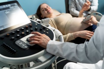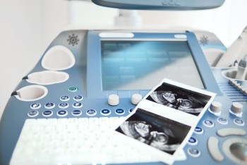
USG Abdomen & Pelvis is conducted to evaluate the structures and organs in the abdominal and pelvic cavities.
Ultrasound / USG Abdomen & Pelvis in India with Cost
USG Abdomen and Pelvis
in Detail
Ultrasound (USG) of the Abdomen and Pelvis is a non-invasive medical imaging technique that employs sound waves to create real-time images of the internal organs within the abdominal and pelvic regions. In this comprehensive guide, we will delve into the purpose, procedure, and significance of USG Abdomen & Pelvis, shedding light on its role in diagnostic medicine.
Understanding USG Abdomen & Pelvis
Purpose of the Scan
USG Abdomen & Pelvis is conducted to evaluate the structures and organs in the abdominal and pelvic cavities. It aids in the diagnosis of various conditions, including but not limited to, liver and kidney diseases, gallbladder issues, reproductive organ abnormalities, and urinary tract problems.
How USG Works
The procedure involves the use of high-frequency sound waves that bounce off internal structures, creating echoes picked up by the ultrasound machine. These echoes are then translated into real-time images, providing a dynamic view of the organs and tissues.
Advantages of USG Abdomen & Pelvis
Non-Invasiveness
One of the key benefits of this imaging technique is its non-invasive nature. Patients experience no exposure to ionizing radiation or discomfort, making it a preferred choice for routine and diagnostic evaluations.
Real-Time Imaging
USG provides immediate, real-time imaging, allowing healthcare professionals to observe organ movements, blood flow, and any abnormalities as they happen. This dynamic aspect is particularly advantageous in assessing the functionality of organs.
Preparing for a USG Abdomen & Pelvis
Fasting and Hydration
In some cases, patients may be required to fast for a few hours before the scan, especially if abdominal organs like the gallbladder need evaluation. Hydration, on the other hand, is usually encouraged to enhance image clarity.
Comfortable Attire
Wearing comfortable and loose-fitting clothing is advisable to facilitate easy access to the abdominal and pelvic areas during the scan. In some instances, patients may be asked to change into a gown provided by the healthcare facility.
The USG Abdomen & Pelvis Procedure
Patient Positioning
Patients lie on an examination table, and a water-based gel is applied to the skin over the abdominal and pelvic areas. The ultrasound transducer is then moved across the skin to capture images.
Transducer Movements
The transducer emits sound waves and captures the returning echoes, creating detailed images displayed on a monitor. The technologist may adjust the transducer's position to examine different areas thoroughly.
Image Interpretation
The real-time images are interpreted by a radiologist or sonographer. They assess the structures and organs, looking for any abnormalities, masses, or changes in blood flow patterns.
Applications of USG Abdomen & Pelvis
Abdominal Organ Evaluation
USG is used to examine organs such as the liver, gallbladder, spleen, pancreas, and kidneys. It helps identify conditions like gallstones, cysts, tumors, or inflammation.
Pelvic Imaging
For pelvic assessments, USG provides insights into the uterus, ovaries, prostate, and bladder. It assists in detecting conditions like fibroids, ovarian cysts, or prostate abnormalities.
Considerations and Limitations
Operator Dependency
The quality of the USG images can be operator-dependent, relying on the skill and experience of the sonographer. However, advancements in technology have minimized this limitation.
Limited View of Certain Structures
USG may have limitations in visualizing structures obscured by gas or bones. In such cases, additional imaging modalities like CT or MRI may be recommended.
Conclusion
In conclusion, USG Abdomen & Pelvis is a versatile and valuable imaging technique in diagnostic medicine. Its non-invasive nature, real-time imaging capabilities, and broad applications make it an essential tool for evaluating and diagnosing a range of abdominal and pelvic conditions.
Frequently Asked Questions (FAQs) for USG Abdomen and Pelvis
1. What is the purpose of a USG Abdomen & Pelvis?
USG Abdomen & Pelvis is performed to evaluate and diagnose conditions in the abdominal and pelvic regions, including organs like the liver, gallbladder, kidneys, uterus, and bladder. It helps in identifying issues such as tumors, cysts, inflammation, and other abnormalities.
2. Is USG Abdomen & Pelvis a painful procedure?
No, USG Abdomen & Pelvis is a non-invasive and painless procedure. It involves the use of sound waves to create images, and patients typically experience no discomfort during the examination.
3. How should I prepare for a USG Abdomen & Pelvis?
Before the scan, you may be required to fast for a few hours, especially if specific abdominal organs need evaluation. It is advisable to wear comfortable clothing and stay hydrated. Your healthcare provider will give you specific instructions tailored to your individual case.
4. Are there any risks associated with USG?
USG is considered a safe imaging technique with no exposure to ionizing radiation. There are generally no known risks or side effects associated with the procedure. It is widely used, even during pregnancy, to assess the health of the fetus.
5. How long does a USG Abdomen & Pelvis take?
The duration of the procedure can vary, but on average, it takes around 30 to 60 minutes. The timing depends on factors such as the complexity of the examination and the areas being evaluated.
6. Can USG detect all abnormalities in the abdominal and pelvic regions?
While USG is a powerful diagnostic tool, it may have limitations in visualizing certain structures, especially those obscured by gas or bones. In some cases, additional imaging modalities like CT or MRI may be recommended for a more comprehensive assessment.
7. Do I need a referral from a doctor to undergo USG Abdomen & Pelvis?
In many cases, a referral from a healthcare provider is required for diagnostic imaging procedures. However, the specific requirements may vary depending on your location and the policies of the healthcare facility.
8. Is USG safe for pregnant women?
Yes, USG is generally considered safe for pregnant women. It does not use ionizing radiation, and the sound waves used during the procedure are not known to harm the fetus. However, it's essential to inform the healthcare provider if you are pregnant or suspect pregnancy before the scan.
9. What information can
USG provide about abdominal organs?
USG can provide detailed information about the size, shape, and texture of abdominal organs such as the liver, gallbladder, spleen, and kidneys. It can help detect conditions like gallstones, liver cysts, and kidney abnormalities.
10. Can USG be used for monitoring ongoing medical conditions?
Yes, USG is often used for monitoring ongoing medical conditions, such as checking the progress of liver disease or assessing the growth and development of a fetus during pregnancy. It allows for real-time observations of changes within the body.
(0)
Login to continue


