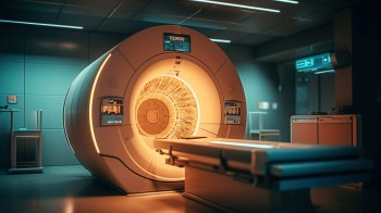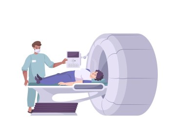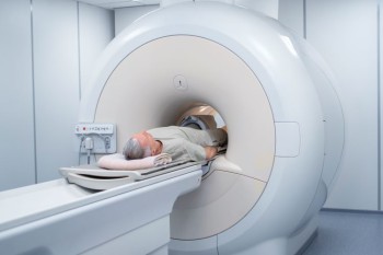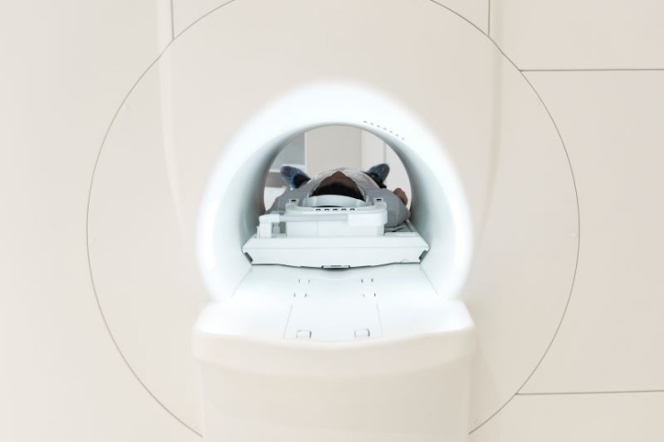
The PSMA PET-CT Scan is a cutting-edge imaging modality that combines the sensitivity of PET with the anatomical detail of CT, targeting the Prostate-Specific Membrane Antigen.
PSMA PET-CT Scan in India with Cost
PSMA PET-CT Scan in
Detail: Revolutionizing Prostate Cancer Imaging for Precision Diagnosis
In the realm of prostate cancer diagnostics, the PSMA PET-CT (Prostate-Specific Membrane Antigen Positron Emission Tomography-Computed Tomography) Scan stands as a state-of-the-art imaging technique, offering unparalleled precision in detecting and evaluating prostate cancer. This comprehensive guide aims to delve into the significance, procedure, and invaluable insights provided by PSMA PET-CT scans, illuminating the advancements in prostate cancer imaging.
Introduction
The PSMA PET-CT Scan is a cutting-edge imaging modality that combines the sensitivity of PET with the anatomical detail of CT, targeting the Prostate-Specific Membrane Antigen. This approach allows for highly accurate localization and characterization of prostate cancer lesions, contributing to enhanced diagnosis, staging, and treatment planning.
Understanding PSMA PET-CT Imaging
PSMA Targeting:
PSMA is a protein overexpressed on the surface of prostate cancer cells. By using radiolabeled compounds that bind specifically to PSMA, the PSMA PET-CT scan allows for the visualization of prostate cancer lesions with remarkable precision.
PET-CT Fusion:
The combination of PET and CT in a single scan provides both functional information about metabolic activity (PET) and detailed anatomical images (CT). This fusion of data enhances the accuracy of lesion localization and characterization.
Importance in Prostate
Cancer Imaging
The PSMA PET-CT Scan has transformative implications in prostate cancer management:
Early Detection: Enables the detection of small or
recurrent prostate cancer lesions with high sensitivity.
Staging Accuracy: Provides accurate staging
information, aiding in the assessment of the extent and spread of the disease.
Treatment Planning: Assists in
planning targeted treatments, such as radiation therapy or surgery, by
precisely identifying cancerous lesions.
Preparation for the Scan
Preparation for a PSMA PET-CT Scan typically involves:
Fasting: Patients may be required to fast
for a few hours before the scan to enhance radiotracer uptake.
Hydration: Adequate hydration is encouraged
to promote optimal radiotracer distribution.
Procedure: Precision in Prostate Cancer
Imaging
Radiotracer Injection: A small amount
of radiotracer, usually Gallium-68 or Fluorine-18, is injected into the
patient's bloodstream.
Uptake Period: The radiotracer circulates through
the body and is selectively taken up by prostate cancer cells expressing PSMA.
Scanning Process: The combined PET-CT scanner
captures both functional and anatomical images, creating a fused image that
highlights PSMA-avid lesions within the prostate and surrounding areas.
Assessment Areas in PSMA
PET-CT Imaging
PSMA PET-CT Scans are employed for various aspects of prostate cancer evaluation, including:
Primary Lesion Detection: Identifying the
primary prostate tumor.
Lymph Node Involvement: Detecting
cancerous involvement in nearby lymph nodes.
Metastatic Spread: Assessing
distant metastases, especially in bones and other organs.
Benefits of PSMA PET-CT
Imaging
Enhanced Sensitivity: Provides superior
sensitivity in detecting prostate cancer lesions, even at low PSA levels.
Accurate Localization: Enables precise
localization and characterization of lesions, guiding targeted interventions.
Treatment Optimization: Facilitates
personalized treatment plans by identifying the extent of the disease.
Risks and Considerations
The risks associated with PSMA PET-CT scans are minimal, primarily related to the injection of radiotracers. The benefits of accurate prostate cancer evaluation generally outweigh the risks.
Clinical Applications
PSMA PET-CT imaging finds applications in various clinical scenarios, including:
Initial Diagnosis: Confirming or
ruling out the presence of prostate cancer.
Staging: Determining the stage of prostate
cancer for appropriate treatment planning.
Recurrence Evaluation: Detecting
recurrent prostate cancer after initial treatment.
Expert Perspectives
Urologists, oncologists, and nuclear medicine specialists collaborate to interpret PSMA PET-CT scans, providing expert insights into prostate cancer diagnosis and management.
Technological Advancements
Ongoing advancements in imaging technology and radiotracer development contribute to the refinement of PSMA PET-CT scans, enhancing sensitivity and specificity.
Patient Experience
While the procedure involves an intravenous injection and lying still during the scan, patients generally find the PSMA PET-CT Scan to be well-tolerated. The benefits of accurate prostate cancer evaluation contribute to informed decision-making.
Conclusion
In conclusion, the PSMA PET-CT Scan emerges as a revolutionary tool in prostate cancer imaging, elevating precision and accuracy in diagnosis and staging. Its applications in early detection, staging accuracy, and treatment planning contribute to improved outcomes and personalized prostate cancer care.
FAQs about PSMA PET-CT Scans:
1. What makes PSMA PET-CT different from traditional imaging for prostate cancer?
PSMA PET-CT combines Positron Emission Tomography (PET) with Computed Tomography (CT) to specifically target prostate cancer cells, providing more accurate and detailed information compared to conventional imaging methods.
2. Is the radiation exposure during a PSMA PET-CT Scan a concern for patients?
The radiation exposure during a PSMA PET-CT Scan is generally considered low and safe. The benefits of accurate prostate cancer detection usually outweigh the associated risks.
3. How long does a PSMA PET-CT Scan take?
The duration of a PSMA PET-CT Scan varies, but the actual imaging process typically takes around 30 to 60 minutes. Additional time may be required for preparation and injection of the radiotracer.
4. Can PSMA PET-CT detect prostate cancer at an early stage?
Yes, one of the significant advantages of PSMA PET-CT is its ability to detect prostate cancer lesions at an early stage, even when the PSA levels are low.
5. Are there any specific patient conditions that might affect the accuracy of the PSMA PET-CT results?
Certain conditions, such as inflammation or recent prostate surgery, may affect PSMA PET-CT results. It's crucial to inform the healthcare team about any relevant medical history.
6. How often is PSMA PET-CT used in prostate cancer follow-up and monitoring?
PSMA PET-CT is often used for follow-up and monitoring after initial prostate cancer treatment to assess treatment effectiveness, detect recurrences, and guide further interventions.
7. Is there any special preparation required before a PSMA PET-CT Scan?
Patients may be advised to fast for a few hours before the scan, and adequate hydration is encouraged. Specific preparation instructions may vary based on individual cases.
8. Can PSMA PET-CT be used for purposes other than prostate cancer detection?
While its primary application is in prostate cancer, PSMA PET-CT may have potential applications in other PSMA-expressing cancers. Research is ongoing to explore its broader utility.
9. How soon can results be expected after a PSMA PET-CT Scan?
Results are typically available shortly after the scan, and the healthcare team will discuss the findings with the patient during a follow-up appointment.
10. Is there an age limit for undergoing a PSMA PET-CT Scan?
No, there is typically no specific age limit for undergoing a PSMA PET-CT Scan. The decision to recommend and perform the scan is based on individual health conditions, clinical indications, and the potential benefits of the imaging procedure. Healthcare providers will assess the overall health status of the patient to ensure the appropriateness and safety of the PSMA PET-CT Scan, regardless of age. It's essential for individuals to discuss their medical history and any concerns with their healthcare team to determine the most suitable diagnostic approach.
These additional FAQs aim to address common queries about PSMA PET-CT Scans, providing a more comprehensive understanding of this advanced imaging technique in the context of prostate cancer diagnosis and management.
(0)
Login to continue
