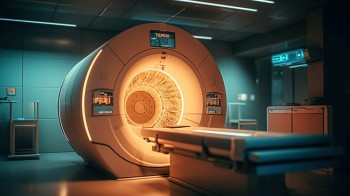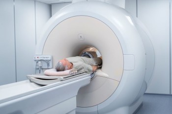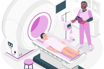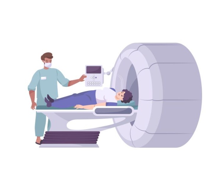
A Brain PET-CT Scan combines two powerful imaging modalities, PET and CT, to offer a comprehensive evaluation of brain structure and function.
Brain PET-CT Scan in India with Cost
Brain PET-CT Scan in
Detail: Illuminating Neurological Insights through Precision Imaging
In the realm of diagnostic imaging, a Brain PET-CT (Positron Emission Tomography-Computed Tomography) Scan stands as a cutting-edge technique, providing unparalleled insights into the intricate details of neurological function and pathology. This comprehensive guide aims to explore the significance, procedure, and invaluable insights provided by Brain PET-CT Scans, offering a detailed understanding of their applications in neuroimaging.
Introduction
A Brain PET-CT Scan combines two powerful imaging modalities, PET and CT, to offer a comprehensive evaluation of brain structure and function. This advanced imaging technique allows for the visualization of both anatomical structures and metabolic activity within the brain, providing critical information for the diagnosis and management of neurological conditions.
Understanding Brain PET-CT Imaging
PET (Positron Emission Tomography): PET involves the injection of a small amount of radioactive material (radiotracer) into the bloodstream. As the radiotracer travels to the brain, it emits positrons, which collide with electrons in the body, producing gamma rays. The PET scanner detects these gamma rays, creating detailed images of metabolic activity within the brain.
CT (Computed Tomography):
CT provides high-resolution anatomical images through X-ray technology. In the context of Brain PET-CT, CT helps precisely localize and align PET images, allowing for a comprehensive assessment of both structure and function.
Importance in
Neuroimaging
The integration of PET and CT in Brain PET-CT Scans offers a holistic approach to neuroimaging. This technique is particularly valuable in:
Oncology: Detecting and staging brain tumors
by assessing both structural abnormalities and metabolic activity.
Neurology: Evaluating neurological conditions
such as epilepsy, Alzheimer's disease, and movement disorders by visualizing
changes in brain metabolism.
Psychiatry: Studying mental health conditions,
including depression and schizophrenia, by observing brain function and
identifying areas of altered activity.
Preparation for the Scan
Preparation for a Brain PET-CT Scan is generally minimal. Patients may be instructed to fast for a few hours before the scan, and it is crucial to inform healthcare providers about any pre-existing medical conditions or medications.
Procedure: Integrating Structure and Function
Assessment
Radiotracer Injection: A small amount
of radiotracer is injected into the patient's bloodstream. The choice of
radiotracer depends on the specific information sought, such as glucose
metabolism or specific neurotransmitter activity.
Uptake Period: After injection, there is a
waiting period during which the radiotracer is absorbed by the brain tissue.
Scanning Process: The combined PET-CT scanner
captures both metabolic activity (PET) and detailed anatomical images (CT)
during a single session. The alignment of these images provides a comprehensive
view of the brain's structure and function.
Assessment Areas in
Brain PET-CT Imaging
Brain PET-CT Scans are employed to assess various neurological aspects, including:
Tumor Detection: Identifying and characterizing
brain tumors based on metabolic activity.
Epilepsy Evaluation: Localizing
areas of abnormal brain activity in patients with epilepsy.
Neurodegenerative Disorders: Observing changes in glucose metabolism associated with conditions like Alzheimer's disease and Parkinson's disease.
Psychiatric Disorders: Investigating
the neural correlates of psychiatric conditions by assessing regional brain
activity.
Benefits of Brain PET-CT
Imaging
Comprehensive Insight: Offers a comprehensive
assessment of both structural and functional aspects of the brain.
Early Detection: Facilitates early detection and
characterization of neurological abnormalities.
Treatment Planning: Assists in
planning and monitoring treatment strategies, especially in oncology and
neurology.
Risks and Considerations
While the radioactive materials used in PET-CT scans expose patients to a small amount of radiation, the benefits of accurate diagnosis and treatment planning generally outweigh the risks. Pregnant women and individuals with certain medical conditions may need specific considerations.
Clinical Applications
Brain PET-CT imaging finds applications in various clinical scenarios, including:
Oncology Departments: Staging and
monitoring brain tumors.
Neurology Clinics: Evaluating and
managing neurological disorders.
Psychiatric Centers: Investigating
the neurobiological basis of psychiatric conditions.
Expert Perspectives
Neuroradiologists, neurologists, and nuclear medicine specialists collaborate to interpret Brain PET-CT scans, providing expert insights into both structural and functional aspects of the brain.
Technological Advancements
Ongoing advancements in imaging technology contribute to the refinement of Brain PET-CT scans, enhancing image resolution, and diagnostic capabilities.
Patient Experience
While the procedure involves minimal discomfort, patients may experience the sensation of intravenous injection and lying still during the scan. The overall experience is well-tolerated, and the benefits of accurate diagnosis often outweigh any temporary inconvenience.
Conclusion
In conclusion, Brain PET-CT Imaging emerges as a revolutionary tool in neuroimaging, seamlessly integrating structural and functional assessments. Its applications in oncology, neurology, and psychiatry contribute to a deeper understanding of neurological conditions, paving the way for precise diagnosis and personalized treatment strategies.
FAQs for Brain PET-CT Scan: Unraveling Neuroimaging Mysteries
1. How long does a Brain PET-CT Scan typically take?
The duration of a Brain PET-CT Scan varies, but it generally takes about 30 to 60 minutes, including the time for radiotracer uptake and the scanning process.
2. Is there any preparation required before a Brain PET-CT Scan?
Patients may be instructed to fast for a few hours before the scan, and it's essential to inform healthcare providers about any pre-existing medical conditions or medications.
3. Does a Brain PET-CT Scan expose patients to a significant amount of radiation?
While there is exposure to a small amount of radiation due to the use of radiotracers, the benefits of accurate diagnosis usually outweigh the risks. Pregnant women and individuals with specific medical conditions may require special considerations.
4. Are there any specific risks associated with Brain PET-CT Scans?
Brain PET-CT Scans are generally safe. However, as with any medical procedure, there are minimal risks. It's crucial to discuss any concerns with the healthcare team beforehand.
5. Can Brain PET-CT Scans detect all types of brain tumors?
Brain PET-CT Scans are powerful tools for detecting and characterizing various types of brain tumors based on their metabolic activity. However, the specific radiotracer used may influence the detection of certain tumor types.
6. Are there any age restrictions for undergoing a Brain PET-CT Scan?
Brain PET-CT Scans can be performed at any age, and there are generally no specific age restrictions. The decision to undergo the scan is based on the patient's overall health and medical needs.
7. How often are Brain PET-CT Scans repeated during cancer treatment?
The frequency of Brain PET-CT Scans during cancer treatment varies based on the specific treatment plan and the response to therapy. The healthcare team will determine the appropriate timing for follow-up scans.
8. Can Brain PET-CT Scans diagnose neurodegenerative disorders like Alzheimer's disease?
Yes, Brain PET-CT Scans can provide valuable insights into neurodegenerative disorders like Alzheimer's disease by assessing changes in glucose metabolism within the brain.
9. Is sedation required for children undergoing Brain PET-CT Scans?
Sedation is generally not required for children undergoing Brain PET-CT Scans. The medical team takes measures to ensure the child's comfort during the procedure.
10. How soon can results be expected after a Brain PET-CT Scan?
Results from a Brain PET-CT Scan are typically available shortly after the scan is completed. The healthcare team will discuss the findings and implications with the patient during a follow-up appointment.
These FAQs provide additional insights into the practical aspects, safety considerations, and applications of Brain PET-CT Scans, addressing common queries that individuals may have before undergoing this advanced neuroimaging technique.
(0)
Login to continue
