The Sacroiliac Joint (SI Joint) is a critical junction in the human skeletal system, connecting the spine to the pelvis.
MRI Sacroiliac (SI) Joint with Cost
MRI Scan Sacroiliac Joint (SI Joint) in Detail: Unlocking the Secrets of
Advanced Imaging
The Sacroiliac Joint (SI Joint) is a critical
junction in the human skeletal system, connecting the spine to the pelvis. To
gain insights into potential issues affecting this joint, healthcare
professionals often turn to Magnetic Resonance Imaging (MRI) for a
comprehensive examination.
The Significance of the Sacroiliac Joint
Anatomy and Function: The SI Joint is formed by the sacrum, the triangular bone at the base of the spine, and the ilium, one of the pelvic bones. It plays a crucial role in supporting the upper body and transferring forces between the spine and the lower limbs.
Common Conditions: Various conditions can affect the SI Joint, leading to pain and discomfort. These include sacroiliitis, arthritis, and ligament sprains. Identifying the root cause is vital for effective treatment.
MRI Examination Process
Preparation: Before the MRI scan, patients may be required to wear a gown and remove any metallic objects. Clear communication with the technologist about any concerns or conditions is crucial.
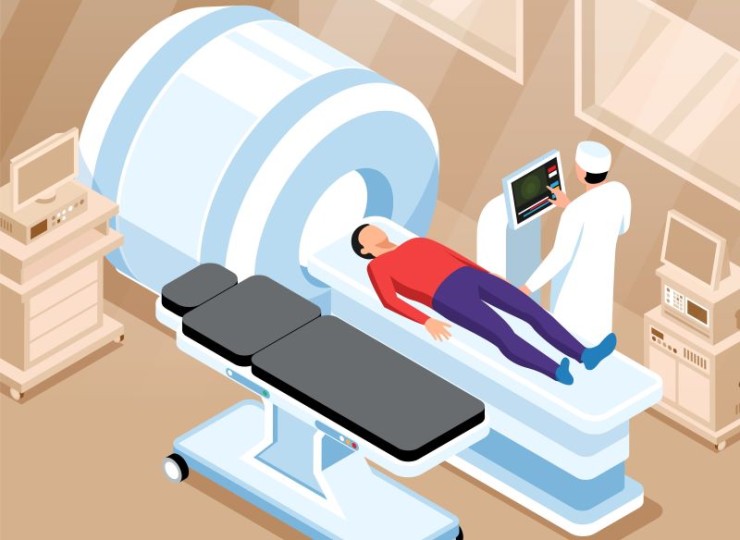
Patient Positioning: Achieving optimal images requires precise patient positioning. Typically, patients lie on their back with the area of interest, the SI Joint, positioned within the MRI machine.
Duration: The MRI examination of the Sacroiliac Joint usually takes around 30 to 60 minutes, during which the patient is required to remain as still as possible.
Advantages of MRI for Sacroiliac Joint Imaging
Soft Tissue Detail: MRI excels in providing detailed images of soft tissues, making it particularly effective for examining the ligaments, cartilage, and other structures around the SI Joint.
Non-Invasiveness: Unlike some diagnostic
procedures, MRI is non-invasive, meaning it does not involve radiation or the insertion of instruments into the body. This makes it a safe option for repeated examinations.
Accuracy: MRI's ability to produce high-resolution images allows healthcare professionals to accurately assess the condition of the Sacroiliac Joint and surrounding structures.
Interpreting MRI Results
Radiologist's Expertise: A radiologist, a medical professional trained in interpreting medical images, reviews the MRI scans. They assess the structures of the SI Joint, looking for abnormalities, inflammation, or other indications of underlying issues.
Consultation: Following the examination, patients typically meet with their healthcare provider to discuss the MRI results. The consultation involves understanding the findings, exploring potential treatment options, and addressing any questions or concerns.
Conclusion
In conclusion, an MRI examination of the Sacroiliac Joint is a valuable diagnostic tool for assessing conditions that may affect this crucial joint. From detailed imaging to accurate diagnosis, MRI plays a pivotal role in the comprehensive care of individuals experiencing SI Joint-related concerns.
FAQs (Frequently Asked Questions) about Sacroiliac (SI) Joint MRI
Q1: Why is an MRI recommended for assessing the
Sacroiliac Joint?
MRI provides detailed images of soft tissues, enabling healthcare professionals to assess ligaments, cartilage, and structures around the Sacroiliac Joint. It's a valuable diagnostic tool for identifying issues like sacroiliitis, arthritis, and ligament sprains.
Q2: Is there any specific preparation required for a Sacroiliac Joint MRI?
Patients may be required to wear a gown and remove metallic objects before the MRI. Clear communication with the technologist about any concerns or conditions is crucial for a successful examination.
Q3: How long does an MRI examination of the Sacroiliac Joint typically take?
The duration of the MRI scan is usually around 30 to 60 minutes. During this time, it's essential for the patient to remain as still as possible to ensure optimal imaging.
Q4: Are there any risks associated with MRI examinations of the Sacroiliac Joint?
MRI is considered a safe and non-invasive procedure, as it does not involve radiation or the insertion of instruments into the body. However, individuals with certain medical conditions or metal implants should inform their healthcare provider.
Q5: Can an MRI detect all issues related to the Sacroiliac Joint?
While MRI provides detailed images, it may not identify every possible issue. The interpretation of results by a skilled radiologist and consultation with a healthcare provider are crucial for a comprehensive understanding of the joint's condition.
Q6: How soon can I expect to receive the results of my Sacroiliac Joint MRI?
The turnaround time for MRI results can vary, but patients typically meet with their healthcare provider shortly after the examination to discuss the findings, explore treatment options, and address any questions or concerns.
Q7: Can I undergo an MRI of the Sacroiliac Joint if I'm pregnant?
MRI is generally considered safe during pregnancy, especially for focused areas like the Sacroiliac Joint. However, it's crucial to inform the healthcare provider about pregnancy to ensure appropriate precautions are taken.
Q8: Is there an age restriction for undergoing a Sacroiliac Joint MRI?
There is no specific age restriction for Sacroiliac Joint MRI scans. The decision to undergo the procedure is based on medical necessity and the recommendation of healthcare professionals.
Q9: Can I bring someone with me during the Sacroiliac Joint MRI scan?
Many healthcare facilities allow a companion to accompany patients during the Sacroiliac Joint MRI scan for support. However, it's advisable to check with the facility beforehand.
(0)
Login to continue


