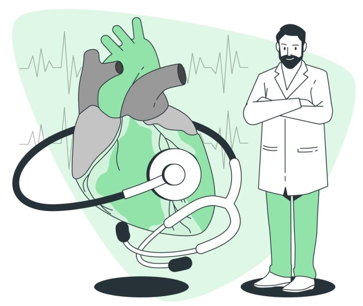
MRI (Magnetic Resonance Imaging) is a state-of-the-art medical imaging technique that offers unparalleled insights into the intricacies of the heart's structure and function.
MRI (Magnetic Resonance
Imaging) is a state-of-the-art medical imaging technique that offers
unparalleled insights into the intricacies of the heart's structure and
function. This non-invasive procedure utilizes a powerful magnetic field and
radio waves to create highly detailed images, providing healthcare
professionals with a comprehensive understanding of the heart's anatomy and
dynamics.
The Procedure:
1.Patient Preparation:
Before the
cardiac MRI scan, patients may need to remove metal objects and inform the
healthcare team about any implants or medical conditions. A contrast agent
might be administered intravenously to enhance image clarity.
2.Positioning:
Patients lie
on a specialized MRI table, which slides into the machine. It's crucial to
remain still during the procedure to ensure the quality of the images.
3.Magnetic Resonance Imaging:
The MRI
machine generates a strong magnetic field, causing hydrogen atoms in the body
to align. When radio waves are applied, these atoms emit signals, captured by
the machine to create detailed cross-sectional images of the heart.
4.Contrast Enhancement:
In some
cases, a contrast agent, usually gadolinium, is injected to highlight specific
areas of the heart, providing additional information for a more accurate
diagnosis.
Significance and
Applications:
1.Diagnostic Precision:
Cardiac MRI
scans excel in diagnosing a myriad of heart conditions, including coronary
artery disease, myocardial infarction, cardiomyopathies, and congenital heart
defects. The high-resolution images aid in identifying subtle abnormalities
that may be missed by other imaging methods.
2.Functional Assessment:
Beyond
structure, cardiac MRI evaluates the heart's function by assessing blood flow,
ventricular volume, and contractility. This functional data is crucial for
understanding the heart's performance and guiding treatment decisions.
3.Treatment Planning:
Cardiac MRI
plays a pivotal role in treatment planning, especially for cardiac surgeries or
interventions. Surgeons rely on these detailed images to strategize procedures
and optimize outcomes.
Advantages:
1.Non-Ionizing Radiation:
Unlike X-rays
or CT scans, cardiac MRI scans do not involve ionizing radiation, making them
safer for repeated use, particularly for patients requiring multiple
assessments.
2.Multi-Parameter Imaging:
Cardiac MRI
provides a comprehensive assessment by combining anatomical and functional data
in a single scan, reducing the need for multiple imaging procedures.
3.Precise Evaluation of Cardiac Tissues:
The high
soft-tissue contrast of MRI enables precise evaluation of cardiac tissues,
contributing to accurate diagnoses and tailored treatment plans.
Conclusion:
In conclusion, a cardiac MRI
scan is a sophisticated and non-invasive tool that revolutionizes cardiac
imaging. From detailed anatomical assessments to functional insights, this
procedure empowers healthcare professionals to deliver precise diagnoses and
personalized care, ultimately enhancing the management of various cardiac
conditions.