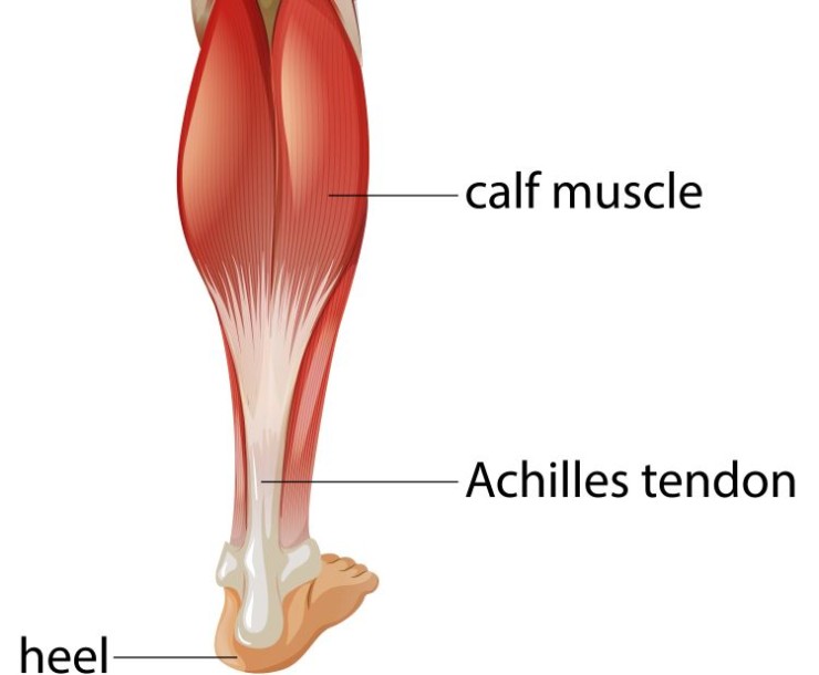
An MRI scan of the calf is a specialized diagnostic imaging procedure aiming to provide detailed insights into the soft tissues of the calf region, encompassing muscles, tendons, ligaments, and surrounding structures.
MRI Scan Calf with Cost
MRI Scan of the Calf: Unraveling the Intricacies of Soft Tissue Imaging
1. Introduction:
An MRI scan of the calf is a specialized diagnostic imaging procedure aiming to provide detailed insights into the soft tissues of the calf region, encompassing muscles, tendons, ligaments, and surrounding structures.
2. Purpose of the Calf MRI Scan:
The primary objective is to diagnose and assess various conditions, including muscle injuries, tendon disorders, ligament tears, and any abnormalities affecting the calf.
3. Patient Preparation:
Patients are generally required to wear comfortable clothing and remove metallic objects before the scan. It is crucial to inform the healthcare team about any medical conditions, allergies, or pregnancy.
4. Imaging Process:
Patient Positioning: During the scan, patients lie on the MRI table, and the calf area is positioned within the scanner.
Contrast Agent: In some cases, a contrast agent may be administered to enhance visibility of specific structures.
5. Duration of the Scan:
The typical duration of an MRI scan of the calf ranges from 30 to 60 minutes. Patient cooperation and remaining still are essential for optimal image quality.
6. Advantages Over Other Imaging Techniques:
Soft Tissue Detail: MRI excels in providing high-resolution images of soft tissues, enabling a thorough evaluation of muscles, tendons, and ligaments.
Non-Invasive Nature: MRI is non-invasive, avoiding exposure to ionizing radiation, making it a safe option for repetitive imaging.
7. Conditions Diagnosed with Calf MRI Scan:
Muscle Injuries: Identification and characterization of muscle strains, tears, or other injuries.
Tendon Disorders: Assessment of tendon health and detection of conditions like Achilles tendonitis.
Ligament Tears: Evaluation of ligaments, crucial for diagnosing conditions affecting calf stability.
8. Patient Experience:
The procedure is generally painless, with minimal discomfort related to the need for immobility during the scan. Open MRI options may be available for individuals with claustrophobia.
9. Interpreting Results:
Results are interpreted by a skilled radiologist who provides detailed insights into the condition of the calf's soft tissues.
A consultation with a healthcare provider follows to discuss findings and plan appropriate treatment.
10. Post-Scan Considerations:
Patients can typically resume normal activities immediately after the MRI calf scan, as there is no recovery time required.
11. Conclusion:
An MRI scan of the calf stands as a crucial diagnostic tool, offering in-depth information for the assessment and treatment of various soft tissue conditions affecting this anatomical region.
12. Future Trends:
Ongoing advancements in MRI technology continue to refine the clarity and specificity of calf imaging, ensuring more accurate diagnoses and treatment planning.
In conclusion, the MRI scan of the calf provides essential insights into the complexities of soft tissue health, aiding in the diagnosis and management of conditions affecting this vital part of the lower limb.
FAQs (Frequently Asked Questions) about Calf MRI
Q1: What is the purpose of an MRI Scan of the Calf?
A1: An MRI Scan of the Calf is performed to provide detailed imaging of the soft tissues in the calf region, including muscles, tendons, ligaments, and surrounding structures. It is primarily used to diagnose conditions such as muscle injuries, tendon disorders, ligament tears, and abnormalities affecting the calf.
Q2: How should I prepare for an MRI Scan of the Calf?
A2: Patients are generally advised to wear comfortable clothing and remove metallic objects. It's crucial to inform the healthcare team about any medical conditions, allergies, or pregnancy. Fasting is usually not required.
Q3: How long does an MRI Scan of the Calf take, and what can I expect during the procedure?
A3: The duration typically ranges from 30 to 60 minutes. Patients lie on the MRI table, and the calf area is positioned within the scanner. Remaining still is essential for optimal image quality. The procedure is generally painless, with minimal discomfort.
Q4: Are there any advantages of an MRI Scan of the Calf over other
imaging techniques?
A4: Yes, MRI provides high-resolution images of soft tissues, enabling a thorough evaluation of muscles, tendons, and ligaments. It is a non-invasive procedure, avoiding exposure to ionizing radiation.
Q5: What conditions can an MRI Scan of the Calf diagnose?
A5: This MRI scan can diagnose various conditions, including muscle injuries (strains or tears), tendon disorders (e.g., Achilles tendonitis), and ligament tears that may affect calf stability.
Q6: Is there any discomfort associated with an MRI Scan of the Calf?
A6: The procedure is generally painless. Minimal discomfort may arise due to the need for immobility during the scan. Open MRI options are available for individuals with claustrophobia.
Q7: How soon can I resume normal activities after an MRI Scan of the Calf?
A7: Patients can typically resume normal activities immediately after the scan, as there is no recovery time required.
Q8: How are the results of an MRI Scan of the Calf interpreted?
A8: Results are interpreted by a skilled radiologist, providing detailed insights into the condition of the calf's soft tissues. A consultation with a healthcare provider follows to discuss findings and plan appropriate treatment.
(0)
Login to continue


