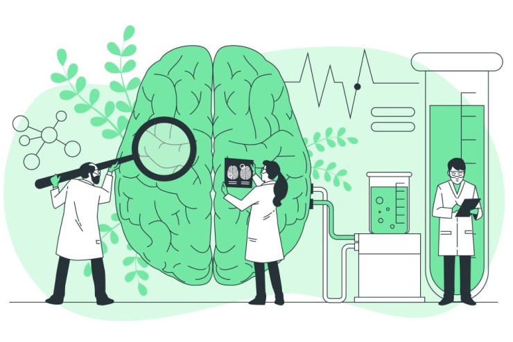
Magnetic Resonance Imaging (MRI) of the brain is a non-invasive medical imaging technique that uses powerful magnets, radio waves, and a computer to generate detailed images of the brain's internal structures.
Magnetic Resonance Imaging
(MRI) of the brain is a non-invasive medical imaging technique that uses
powerful magnets, radio waves, and a computer to generate detailed images of
the brain's internal structures. It is a valuable tool for diagnosing and evaluating
a wide range of neurological conditions. Here's a detailed overview of an MRI
brain scan:
1.Purpose:
·Diagnostic Imaging: The primary purpose of an MRI brain scan is to
provide detailed and high-resolution images of the brain to aid in the
diagnosis and evaluation of neurological conditions.
2.Indications:
·Headaches:
To investigate the cause of persistent or severe headaches.
·Seizures:
For assessing the structural causes of seizures.
·Stroke:
To detect and evaluate the extent of ischemic or hemorrhagic strokes.
·Tumors:
To identify and characterize brain tumors.
·Trauma:
Following head injuries to assess for any structural damage.
·Neurological Disorders: For diagnosing conditions such as multiple
sclerosis, Alzheimer's disease, or other neurodegenerative disorders.
·Vascular Abnormalities: To detect abnormalities in blood vessels, such
as aneurysms or arteriovenous malformations (AVMs).
3.Technique:
·Magnetic Resonance Imaging (MRI): The imaging technique relies on the interaction
of hydrogen atoms in the body with a strong magnetic field.
·Pulse Sequences: Different pulse sequences, including T1-weighted, T2-weighted, and
FLAIR (fluid-attenuated inversion recovery), provide information about various
tissue characteristics.
·Contrast-Enhanced Imaging: In some cases, a gadolinium-based contrast
agent may be administered to enhance the visibility of certain structures or
abnormalities.
4.Procedure:
·Patient Preparation: Patients typically do not require special
preparations, but they may be asked to remove metal objects and inform the
healthcare provider of any implants or conditions that may affect the scan.
·Positioning: The patient lies on a moveable bed that enters the MRI scanner. It is
essential to remain still during the scan to ensure clear images.
·Duration:
The procedure usually takes 30 to 60 minutes, during which multiple image
slices are acquired.
5.Interpretation:
·Radiologist's Assessment: The acquired images are interpreted by a
radiologist, who assesses the brain's structures for any abnormalities,
lesions, or pathological changes.
6.Clinical Significance:
·Disease Diagnosis: MRI brain scans are instrumental in diagnosing
a wide range of neurological conditions, including tumors, strokes, and
degenerative disorders.
·Treatment Planning: Results from the MRI scan guide healthcare
providers in planning appropriate treatments based on the identified
abnormalities.
·Monitoring:
Serial MRI scans may be used to monitor the progression or response to
treatment in certain conditions.
7.Limitations:
·Contrast Agents: Some patients may have contraindications to contrast agents, and
alternative imaging approaches may be considered.
·Claustrophobia: Patients with claustrophobia may find the confined space of the MRI
scanner challenging.
Overall, MRI brain scans
provide detailed anatomical information, aiding healthcare professionals in
diagnosing and managing various neurological conditions. The versatility of MRI
allows for the visualization of soft tissues, blood vessels, and abnormalities
with high precision.