Magnetic Resonance Imaging (MRI) of the brain with a focus on the pituitary gland is a specialized imaging technique used to visualize the pituitary gland, a small, pea-sized gland located at the base of the brain.
Magnetic Resonance Imaging
(MRI) of the brain with a focus on the pituitary gland is a specialized imaging
technique used to visualize the pituitary gland, a small, pea-sized gland
located at the base of the brain. This imaging is valuable for detecting and
evaluating various pituitary disorders. Here's a detailed overview:
1.Purpose:
·Pituitary Assessment: The primary purpose of MRI brain with pituitary
is to assess the anatomy and function of the pituitary gland.
·Detection of Abnormalities: It is used to detect and evaluate abnormalities
such as tumors, cysts, hemorrhages, and other lesions affecting the pituitary
gland.
2.Indications:
·Hormonal Imbalances: When patients present with hormonal imbalances
or symptoms suggestive of pituitary dysfunction.
·Visual Disturbances: In cases where there are visual disturbances or
symptoms related to compression of surrounding structures by the pituitary
mass.
·Headaches:
When headaches or neurological symptoms are suspected to be associated with
pituitary abnormalities.
3.Technique:
·Structural Imaging: The MRI component involves obtaining detailed
structural images of the brain using various sequences such as T1-weighted,
T2-weighted, and gadolinium-enhanced sequences.
·Contrast-Enhanced Imaging: In many cases, a gadolinium-based contrast
agent is administered to enhance the visibility of pituitary lesions and
surrounding structures.
4.Procedure:
·Patient Preparation: Patients undergoing MRI brain with pituitary
imaging generally do not require special preparations. However, it is crucial
to inform healthcare providers about any pre-existing conditions, allergies, or
medications.
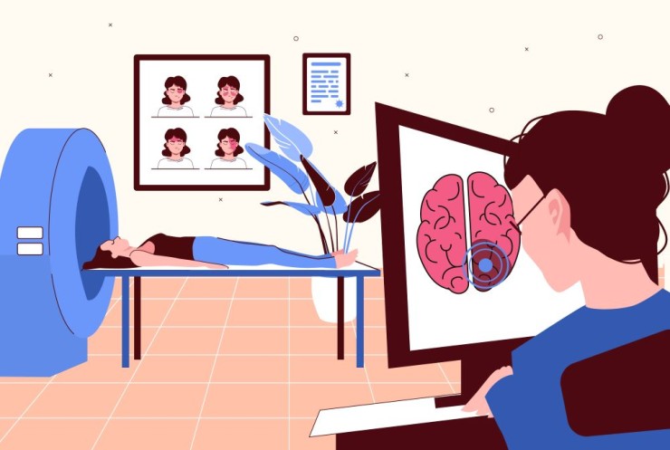
·Image Acquisition: The patient is positioned within the MRI
scanner, and images are acquired during the imaging session, with a specific
focus on the pituitary region.
5.Interpretation:
·Radiologist's Assessment: The acquired images are interpreted by a
radiologist, who assesses the size, shape, and signal characteristics of the
pituitary gland.
·Lesion Detection: The radiologist looks for abnormalities such as
tumors, cysts, or other lesions affecting the pituitary gland.
6.Clinical Significance:
·Tumor Identification: MRI of the pituitary is crucial for identifying
and characterizing pituitary tumors, which can be functional (secreting
hormones) or non-functional.
·Assessment of Surrounding Structures: The imaging helps evaluate the impact of
pituitary lesions on nearby structures, including the optic chiasm, which can
lead to visual disturbances if compressed.
·Treatment Planning: Findings from the MRI assist in treatment
planning, whether it involves medical management, surgery, or other
interventions.
7.Limitations:
·Small Lesion Detection: While MRI is highly sensitive, very small
lesions or those with subtle characteristics may require additional imaging or
follow-up.
·Functional Assessment: MRI primarily provides anatomical information,
and functional assessment may require additional tests, such as hormonal
assays.
MRI of the brain with a focus
on the pituitary is a valuable tool in the diagnosis and management of
pituitary disorders. It provides detailed information that guides clinicians in
understanding the nature of pituitary lesions and formulating appropriate
treatment plans.