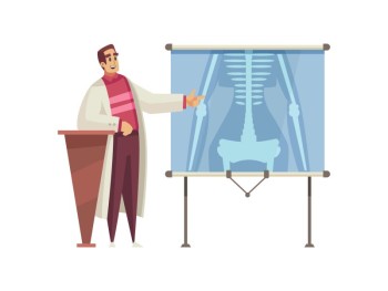
A Chest X-ray is a common diagnostic imaging procedure that uses X-ray technology to visualize the structures within the chest, including the lungs, heart, blood vessels, and bones.
Chest X-ray with Cost
Chest X-ray in Detail
Introduction
A Chest X-ray is a common diagnostic imaging procedure that uses X-ray technology to visualize the structures within the chest, including the lungs, heart, blood vessels, and bones.
Purpose and Procedure
The primary purpose of a Chest X-ray is to assess and diagnose conditions affecting the chest and respiratory system. During the procedure, the patient is positioned in front of the X-ray machine, and X-ray images are captured. The radiographer may give specific instructions on breath-holding to ensure clear images.
Types of Chest X-rays
There are various types of Chest X-rays, each serving different purposes. Routine Chest X-rays provide an overall view of the chest, while Portable Chest X-rays are performed at the bedside for patients who cannot move easily. Specialized Chest X-rays, such as CT Angiography and Fluoroscopy, offer detailed imaging for specific diagnostic needs.
Indications and Contraindications
Chest X-rays are indicated for a range of medical conditions, including pneumonia, lung infections, fractures, and heart conditions. Contraindications are minimal, and the benefits of the diagnostic information often outweigh any potential risks. Pregnant individuals are usually advised to inform their healthcare provider before the procedure.
Risks and Safety Measures
The risks associated with Chest X-rays are minimal, as the amount of radiation exposure is relatively low. Healthcare professionals take precautions to minimize radiation exposure, especially in sensitive populations. Lead aprons may be used to protect areas of the body not being imaged.
Interpretation of Results
Interpreting Chest X-ray results requires expertise, typically provided by radiologists and healthcare providers. Abnormalities such as lung nodules, fractures, or signs of infections can be identified through careful analysis of the X-ray images.
Comparison with Alternative Diagnostic Methods
Chest X-rays are often compared with other imaging techniques, such as CT scans and MRI, to determine the most appropriate diagnostic approach for specific cases. Each method has its advantages and limitations, and the choice depends on the clinical context.
Technological Advancements in Chest X-ray Imaging
Recent technological advancements in Chest X-ray imaging include the adoption of digital radiography, enhancing image quality and allowing for easier storage and sharing of images. Artificial intelligence (AI) integration has also shown promise in aiding radiologists in image interpretation.
Common Misconceptions
Addressing common misconceptions about Chest X-rays, such as concerns about radiation exposure or the invasiveness of the procedure, is important for ensuring informed decision-making and alleviating patient anxiety.
Future Trends and
Developments
The future of Chest X-ray imaging holds exciting possibilities, with ongoing advancements expected to enhance diagnostic precision and patient comfort. Continued improvements in technology may lead to faster imaging processes and reduced radiation exposure.
Patient Experiences and Testimonials
Personal stories from individuals who have undergone Chest X-rays provide valuable insights into the real-world impact of the procedure. These testimonials offer a human perspective on the patient experience and its role in their diagnostic journey and subsequent treatment.
Conclusion
In conclusion, Chest X-ray remains a fundamental tool in diagnostic imaging, providing crucial information for healthcare professionals. Its versatility, coupled with technological advancements, makes it a cornerstone in the assessment of chest and respiratory conditions.
FAQs (Frequently Asked Questions) about Chest X-ray
Is a Chest X-ray painful?
No, a Chest X-ray is a non-invasive procedure, and patients typically experience no pain. The brief exposure to radiation is minimal.
How long does a Chest X-ray procedure take?
The procedure itself usually takes a few minutes. The entire process, including preparation and imaging, may take around 15 to 30 minutes.
Can Chest X-rays detect lung cancer?
Chest X-rays may identify suspicious lesions, but further imaging such as CT scans is often needed for a definitive diagnosis of lung cancer.
Are there any side effects from a Chest X-ray?
There are typically no immediate side effects. In rare cases, some individuals may experience a mild skin reaction at the site of radiation exposure.
Is a Chest X-ray safe during pregnancy?
While the radiation exposure is low, pregnant individuals are advised to inform their healthcare provider before undergoing a Chest X-ray to discuss potential risks and benefits.
Can a Chest X-ray detect conditions other than respiratory issues?
Yes, a Chest X-ray can also identify conditions affecting the heart, blood vessels, bones, and surrounding structures within the chest.
How often should individuals undergo routine Chest X-rays for preventive
healthcare?
The frequency of routine Chest X-rays depends on individual health factors and medical history. Healthcare providers will recommend them as needed.
Is there any preparation required before a Chest X-ray?
Generally, no special preparation is needed for a routine Chest X-ray. However, individuals may be asked to remove jewelry or other metal objects.
Can Chest X-rays be performed on pediatric patients?
Yes, Chest X-rays are commonly used in pediatric cases to assess respiratory and chest conditions in children.
What is the difference between Chest X-ray and CT Chest Scan?
While both imaging methods capture images of the chest, CT Chest Scans provide more detailed and cross-sectional images, often used for specific diagnostic purposes.
(0)
Login to continue


