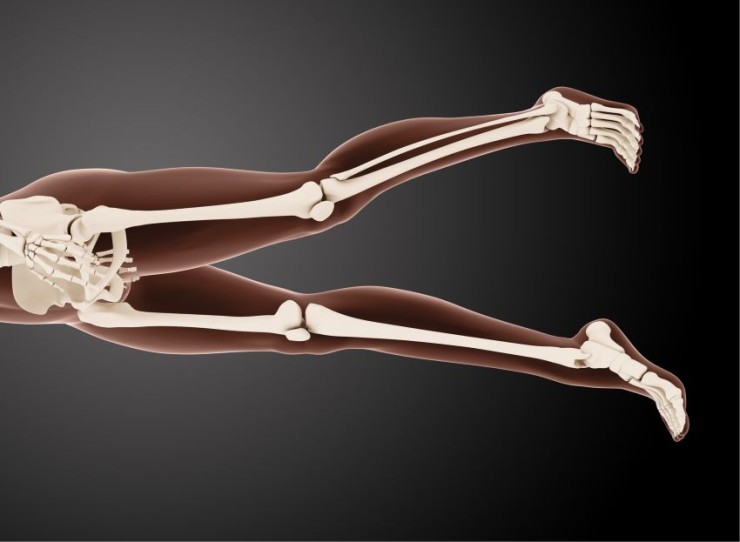
Scanogram X-ray, a crucial component of skeletal imaging, plays a pivotal role in assessing bone structures and identifying abnormalities.
Scanogram X-ray with Cost
Scanogram X-ray in Detail
Introduction
Scanogram X-ray, a crucial component of skeletal
imaging, plays a pivotal role in assessing bone structures and identifying
abnormalities. This diagnostic technique provides valuable insights into limb
length, alignment, and overall skeletal health.
History of Scanogram X-ray
The
history of Scanogram X-ray is marked by continuous advancements in imaging
technology. From its early days to the present, the technique has undergone
significant evolution, enhancing its diagnostic capabilities for skeletal
assessments.
Purpose of Scanogram
X-ray
The primary purpose of Scanogram X-ray is to detect
abnormalities in bone structures. It proves particularly effective in
evaluating limb length discrepancies, assessing alignment issues, and providing
essential information for orthopedic interventions.
Types of Scanogram X-rays
Scanogram X-rays are versatile and come in various
types, each tailored to examine specific aspects of the skeletal system.
Long-Leg Scanogram, Pelvic Scanogram, and Spine Scanogram offer distinct
perspectives, aiding in comprehensive skeletal diagnostics.
Procedure of Scanogram X-ray
Before
undergoing a Scanogram X-ray, patients undergo specific preparations to ensure
optimal imaging results. The procedure involves the careful positioning of the
patient and the acquisition of detailed images to visualize the skeletal
structures of interest.
Indications and
Contraindications
Scanogram X-ray is recommended in cases where
abnormalities or issues related to bone structures are suspected. However,
healthcare providers may avoid the procedure in certain instances, such as
during pregnancy or when alternative diagnostic methods are more suitable.
Risks and Safety Measures
While
generally safe, Scanogram X-ray does carry some inherent risks, including
exposure to ionizing radiation. Healthcare professionals take necessary
precautions, such as using minimal radiation doses, to minimize these risks and
ensure patient safety.
Interpreting Scanogram
X-ray Results
Interpreting Scanogram X-ray results requires a
thorough understanding of normal and abnormal findings within the skeletal
system. Healthcare professionals analyze the images to identify any structural
or alignment abnormalities that may impact a patient's musculoskeletal health.
Comparison with Other Diagnostic Techniques
Comparing
Scanogram X-ray with alternative diagnostic methods, such as CT scans or MRI,
highlights the unique advantages and limitations of each approach.
Understanding these differences assists medical professionals in choosing the
most suitable imaging technique for specific skeletal assessments.
Case Studies
Real-life case studies provide tangible examples of
how Scanogram X-ray has played a pivotal role in diagnosing and treating
various skeletal conditions. These cases illustrate the practical application
and success of the imaging technique in diverse orthopedic scenarios.
Technological Advancements in Scanogram X-ray
Recent
advancements in imaging technology have significantly improved Scanogram X-ray,
making it more efficient and patient-friendly. Innovations in equipment
contribute to enhanced accuracy and efficiency, providing valuable information
for orthopedic diagnoses and interventions.
Common Misconceptions
about Scanogram X-ray
Addressing common myths and misconceptions
surrounding Scanogram X-ray is crucial for fostering confidence in individuals
scheduled for the procedure. Clearing up misunderstandings ensures informed
decision-making and reduces anxiety associated with skeletal imaging.
Future Trends and Developments
As
technology continues to advance, the future of Scanogram X-ray holds promising
possibilities. Emerging trends hint at increased diagnostic accuracy, reduced
invasiveness, and enhanced patient comfort in skeletal imaging, paving the way
for more personalized orthopedic care.
Patient Experiences and Testimonials
Personal stories from
individuals who have undergone Scanogram X-ray provide valuable insights into
the real-world impact of the procedure. These testimonials offer a human
perspective on the patient experience and its role in their orthopedic journey.
Conclusion
In
conclusion, Scanogram X-ray emerges as a vital tool in skeletal diagnostics,
providing essential information for healthcare professionals and orthopedic
specialists. Its rich history, diverse applications, and ongoing technological
improvements make it an indispensable component of modern musculoskeletal
imaging.
FAQs (Frequently Asked Questions) about Scanogram X-ray
Is Scanogram X-ray suitable for pediatric patients?
Yes, Scanogram X-ray can be adapted for pediatric patients, providing valuable insights into their skeletal health and development.
Can Scanogram X-ray detect joint-related issues?
While primarily focused
on bone structures, Scanogram X-ray can contribute to identifying certain
joint-related issues affecting the alignment and function of bones.
How often is Scanogram X-ray recommended for orthopedic assessments?
The frequency of Scanogram X-ray assessments depends on individual patient needs and the specific orthopedic conditions being monitored.
Does Scanogram X-ray require any specific post-procedure care?
In most cases, individuals can resume their normal activities after the procedure. However, specific post-procedure care instructions may be provided by healthcare professionals based on individual cases.
Can Scanogram X-ray be used for assessing spinal conditions?
Yes, Spine Scanogram is a specific type of Scanogram X-ray that focuses on assessing the vertebral column, making it valuable for detecting spinal abnormalities and alignment issues.
Can Scanogram X-ray detect congenital skeletal abnormalities?
Yes, Scanogram X-ray is valuable in identifying congenital skeletal abnormalities, offering insights into the structural development of bones from birth.
Is Scanogram X-ray suitable for assessing joint replacement success?
Scanogram X-ray can be utilized to assess the success and alignment of joint replacement surgeries, providing essential information for postoperative care.
Are there any age limitations for undergoing a Scanogram X-ray?
Scanogram X-ray is generally suitable for individuals of all ages. However, healthcare providers may consider alternative imaging methods for very young or elderly patients based on specific medical conditions.
Can Scanogram X-ray be repeated if needed for ongoing monitoring?
Yes, Scanogram X-ray can be repeated as needed for ongoing monitoring of skeletal conditions. The frequency of repeat scans is determined by the healthcare provider based on the patient's individual circumstances.
What information can a Long-Leg Scanogram provide that other types may
not?
A Long-Leg Scanogram is specifically designed to assess limb length and alignment over an extended area, making it particularly useful for detecting discrepancies or abnormalities in the entire lower extremities.
(0)
Login to continue


