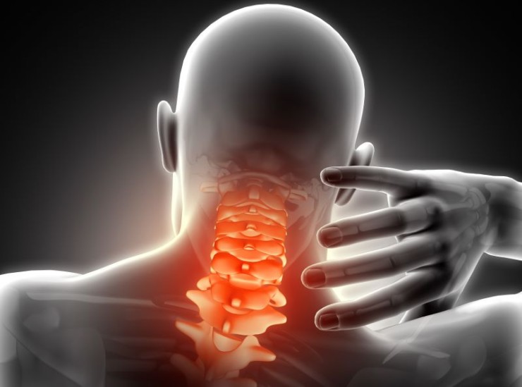
Cervical spine X-rays, specifically the Anteroposterior (AP) and Lateral (Lat) views, are invaluable diagnostic tools used to assess the health and structure of the neck's vertebrae.
Cervical Spine X-ray AP & LAT View with Cost
Exploring Cervical Spine
X-ray: Understanding AP & Lat Views
Introduction
Cervical spine X-rays, specifically the Anteroposterior (AP) and Lateral (Lat) views, are invaluable diagnostic tools used to assess the health and structure of the neck's vertebrae. In this comprehensive guide, we'll delve into the purpose, preparation, and interpretation of these essential imaging techniques.
Purpose of Cervical
Spine X-rays
Cervical spine X-rays serve a vital role in diagnosing and evaluating various conditions affecting the neck and upper spine. Whether investigating chronic neck pain, trauma, or preparing for surgery, these X-rays provide detailed insights into the cervical anatomy.
Preparation for the X-ray
Patient preparation is crucial for obtaining clear and accurate images. The patient is positioned facing the X-ray source for AP views and laterally for Lat views. Radiographers take precautions to ensure patient comfort and adherence to proper positioning.
Understanding AP Views
Positioning and Technique
In Anteroposterior views, the patient faces the X-ray source while the neck is extended and centered. This frontal perspective captures the entire cervical spine, allowing for a comprehensive analysis.
Key Details Captured
AP views reveal the alignment of vertebral bodies, intervertebral spaces, and the presence of any abnormalities such as fractures, dislocations, or degenerative changes.
Interpretation of AP
Views
Radiologists analyze AP views to distinguish normal from abnormal findings. Conditions like cervical spondylosis, herniated discs, or spinal stenosis become apparent through careful examination of these images.
Significance of Lat Views
Positioning and Technique
In Lateral views, the patient is positioned laterally to the X-ray source. This side perspective provides valuable information about the cervical spine's alignment and the condition of individual vertebrae.
Advantages in Diagnosis
Lat views are particularly useful in detecting fractures, assessing curvature abnormalities, and identifying issues like lordosis or kyphosis that might not be as visible in AP views.
Analyzing Lat Views
Healthcare providers carefully analyze lateral perspectives to gain a comprehensive understanding of the cervical spine's health. Issues such as lordosis or kyphosis, which might not be evident in AP views alone, become apparent when assessing lateral X-rays.
When Cervical Spine
X-rays are Recommended
Cervical spine X-rays are recommended in various medical scenarios, including:
Chronic neck pain
Traumatic injuries
Preoperative assessments
These X-rays aid healthcare professionals in formulating accurate diagnoses and effective treatment plans.
Safety Measures During Cervical Spine X-rays
Acknowledging concerns about radiation exposure, safety measures are implemented during cervical spine X-rays. These include radiation dosage, utilizing lead shielding, and ensuring patient comfort throughout the procedure.
Comparison with Other Diagnostic Tools
While cervical spine X-rays offer valuable insights, they are not the sole diagnostic tool available. Comparisons with Magnetic Resonance Imaging (MRI) and Computed Tomography (CT) scans highlight the strengths and limitations of each method, guiding healthcare professionals in choosing the most appropriate imaging modality for a given situation.
Role in Injury Assessment
In cases of trauma or injury, cervical spine X-rays become integral in assessing the extent of damage. Detecting fractures, dislocations, and identifying potential spinal cord compression are crucial steps in formulating an effective treatment plan.
Common Challenges in
Interpretation
Interpreting cervical spine X-rays is not without its challenges. Factors such as patient movement, overlapping structures, or pre-existing conditions may hinder clarity. However, with advancements in technology and enhanced imaging techniques, these challenges can be mitigated.
Future Developments in Cervical Imaging
The landscape of medical imaging is ever-evolving, and cervical spine imaging is no exception. Ongoing advancements in technology promise improvements in diagnostic accuracy, reducing the likelihood of false positives or negatives.
Patient Experience and Feedback
Understanding the patient's perspective is vital in healthcare. Sharing real-life experiences and patient testimonials can alleviate concerns and demystify the process of undergoing cervical spine X-rays. Creating a patient-friendly environment contributes to a positive healthcare journey.
Conclusion
In conclusion, cervical spine X-rays, particularly the AP and Lat views, are invaluable tools in the realm of diagnostic imaging. From identifying structural abnormalities to guiding surgical interventions, these imaging techniques provide crucial information for healthcare professionals. As technology continues to advance, the future holds even more promise for enhancing diagnostic accuracy and patient outcomes.
FAQs (Frequently Asked Questions)
Are cervical spine X-rays painful?
No, cervical spine X-rays are non-invasive and painless. Patients might need to hold specific positions during the procedure, but discomfort is minimal.
How long does it take to get the results of a cervical spine X-ray?
Results are typically available shortly after the imaging procedure, and healthcare providers will discuss them with the patient during a follow-up appointment.
Can cervical spine X-rays be performed during pregnancy?
While precautions are taken to minimize radiation exposure, pregnant individuals should inform their healthcare provider before undergoing X-rays.
Are there alternatives to cervical spine X-rays?
Yes, alternatives such as MRI and CT scans are available, each with its own set of advantages and limitations.
What conditions can be detected through cervical spine X-rays?
Cervical spine X-rays can detect conditions such as fractures, herniated discs, spinal stenosis, and alignment issues.
How often are cervical spine X-rays recommended?
The frequency of X-rays depends on the individual's medical condition. They are typically done as needed for diagnosis or follow-up assessments.
Can cervical spine X-rays detect nerve-related issues?
While X-rays primarily capture bone structures, they can indirectly indicate nerve-related issues by revealing abnormalities or compression in the spinal column.
Are there age restrictions for undergoing cervical spine X-rays?
Cervical spine X-rays can be performed at any age, and there's no specific age restriction. The necessity is determined based on clinical indications.
Do cervical spine X-rays expose patients to a significant amount of
radiation?
The amount of radiation in X-rays is minimal, and healthcare providers take precautions to ensure exposure is as low as reasonably achievable while still obtaining necessary diagnostic information.
Can cervical spine X-rays diagnose conditions like arthritis?
Yes, cervical spine X-rays can help diagnose arthritis by revealing joint space narrowing, bone spurs, and other characteristic signs of the condition.
What should I expect during a cervical spine X-ray procedure?
The procedure is quick and painless. You will be asked to hold specific positions for a short duration while the X-ray machine captures images.
Can cervical spine X-rays be repeated if needed?
Yes, if further evaluation or follow-up is required, healthcare providers may recommend additional X-rays to monitor changes or assess treatment effectiveness.
Are cervical spine X-rays covered by insurance?
In most cases, insurance covers medically necessary X-rays. It's advisable to check with your insurance provider to confirm coverage details.
How can I alleviate anxiety before a cervical spine X-ray?
Communicate any concerns with the healthcare team beforehand. They can provide information, address fears, and make the experience more comfortable for you.
(0)
Login to continue


