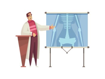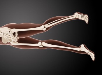
Knee Joint Anteroposterior (AP) and Lateral X-rays are diagnostic imaging procedures that provide valuable insights into the structure and health of the knee joint.
Both Knee Joint AP & Lateral X-ray with Cost
Knee Joint AP & Lateral X-ray in Detail
Introduction
Knee Joint Anteroposterior (AP) and Lateral X-rays are diagnostic imaging procedures that provide valuable insights into the structure and health of the knee joint. These X-rays capture images from two different perspectives, offering a comprehensive assessment.
Purpose and Procedure
The primary purpose of Knee Joint AP and Lateral X-rays is to evaluate the bones, joints, and soft tissues of the knee. During the procedure, the patient is positioned differently for each view. The AP view captures an image from the front, while the lateral view captures a side profile, ensuring a thorough examination of the entire knee joint.
Types of Knee Joint X-rays
Anteroposterior (AP) View: This view captures the knee joint from the front, providing a detailed image of the patella, femur, tibia, and fibula.
Lateral View: This view captures the knee joint from the side, allowing assessment of the joint space, alignment, and any abnormalities.
Indications and Contraindications
Knee Joint X-rays are indicated for various conditions, including fractures, arthritis, ligament injuries, and alignment issues. Contraindications are minimal, and the benefits of obtaining crucial diagnostic information often outweigh any potential risks.
Risks and Safety Measures
The risks associated with Knee Joint X-rays are minimal, as the exposure to radiation is limited. Healthcare professionals take precautions to minimize radiation exposure, particularly in sensitive populations. Lead shielding may be used to protect areas not being imaged.
Interpretation of Results
Interpreting Knee Joint X-ray results requires expertise, typically provided by radiologists and healthcare providers. Abnormalities such as fractures, dislocations, or signs of arthritis can be identified through careful analysis of the X-ray images.
Comparison with Alternative Diagnostic Methods
Knee Joint X-rays are often compared with other imaging techniques, such as MRI and CT scans. While X-rays provide a quick and cost-effective initial assessment, more advanced imaging methods may be employed for detailed evaluation in certain cases.
Technological Advancements in Knee Joint X-ray Imaging
Recent technological advancements in Knee Joint X-ray imaging include the adoption of digital radiography, enhancing image quality and allowing for easier storage and sharing of images. These advancements contribute to a more detailed and precise assessment of knee joint conditions.
Common Misconceptions
Addressing common misconceptions about Knee Joint X-rays, such as concerns about radiation exposure or discomfort during the procedure, is crucial for ensuring informed decision-making and alleviating patient anxiety.
Future Trends and Developments
The future of Knee Joint X-ray imaging holds promising possibilities, with ongoing advancements expected to enhance diagnostic precision and patient comfort. Continued improvements in technology may lead to faster imaging processes and further reduction of radiation exposure.
Patient Experiences and Testimonials
Personal stories from individuals who have undergone Knee Joint X-rays provide valuable insights into the real-world impact of the procedure. These testimonials offer a human perspective on the patient experience and its role in their knee joint diagnostic journey.
Conclusion
In conclusion, Knee Joint AP and Lateral X-rays play a pivotal role in assessing knee joint conditions, providing essential information for healthcare professionals. Their versatility, coupled with technological advancements, makes them integral components of modern orthopedic diagnostics.
FAQs (Frequently Asked Questions) about Both Knee Joint AP and LAT X-ray
Is the positioning uncomfortable during Knee Joint X-rays?
While specific
positioning is necessary, it is generally well-tolerated, and radiographers
ensure patient comfort throughout the procedure.
How long does it take to obtain both Knee Joint AP and Lateral X-ray images?
The
entire process, including preparation and imaging, usually takes around 15 to
30 minutes.
Can Knee Joint X-rays detect cartilage injuries?
While X-rays primarily visualize bones, they may reveal indirect signs of cartilage injuries. For detailed assessment, additional imaging methods like MRI may be recommended.
Are Knee Joint X-rays safe during pregnancy?
The radiation exposure is minimal, and the benefits of obtaining diagnostic information often outweigh potential risks. However, pregnant individuals should inform their healthcare provider before the procedure.
How often should individuals undergo Knee Joint X-rays for preventive
orthopedic care?
The frequency of Knee Joint X-rays depends on individual health factors, symptoms, and the recommendations of healthcare providers. Routine X-rays are not typically performed unless medically indicated.
Can Knee Joint X-rays identify meniscus tears?
While X-rays primarily visualize bones, they may reveal indirect signs of meniscus injuries, such as joint space narrowing. However, for detailed assessment, additional imaging methods like MRI may be recommended.
Is there any age limit for undergoing Knee Joint X-rays?
Knee Joint X-rays can be performed at any age, and the decision is based on clinical indications rather than age. Healthcare providers assess the necessity based on individual health conditions.
Can Knee Joint X-rays be performed on individuals with metal implants?
Yes, Knee Joint X-rays can be performed on individuals with metal implants, but it's essential to inform healthcare providers about any metallic objects in the body to adjust imaging parameters if needed.
What conditions can Knee Joint X-rays help diagnose?
Knee Joint X-rays are valuable for diagnosing various conditions, including fractures, arthritis, ligament injuries, and joint alignment issues.
Is sedation required for children undergoing Knee Joint X-rays?
Sedation is typically not required for Knee Joint X-rays in children, as the procedure is quick and non-invasive. Radiographers ensure a comfortable and child-friendly environment.
(0)
Login to continue


