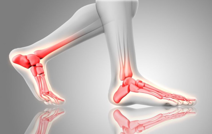
X-ray imaging plays a pivotal role in the realm of medical diagnostics, particularly when examining intricate areas like the ankle, foot, and heel.
X-ray Ankle or
Foot or Heel joint AP and LAT View with Cost
Introduction
X-ray imaging plays a pivotal role in the realm of medical diagnostics, particularly when examining intricate areas like the ankle, foot, and heel. Through the lens of both anterior-posterior (Ap) and lateral (Lat) views, X-rays offer a detailed glimpse into the skeletal landscape, aiding healthcare professionals in accurate diagnoses and treatment plans.
Understanding X-ray Views
Before delving into the specifics of ankle, foot, and heel X-rays, it's crucial to comprehend the significance of Ap and Lat views. The Ap view provides a frontal perspective, while the Lat view captures the side angle. Each view contributes unique information, collectively offering a comprehensive understanding of the structures under examination.
Preparation for X-ray Imaging
Patient preparation is a vital aspect of ensuring the success of an X-ray procedure. Clear communication about the necessity of removing jewelry or accessories, wearing appropriate attire, and following safety protocols creates a conducive environment for obtaining accurate images. Safety measures, including lead aprons and thyroid shields, are employed to minimize radiation exposure.
Anatomy of the Ankle, Foot, and Heel
To appreciate the diagnostic capabilities of ankle, foot, and heel X-rays, a basic understanding of the anatomy is essential. The intricate network of bones, joints, and soft tissues in these areas demands a nuanced approach to imaging. The examination focuses on specific areas of interest, offering a targeted diagnostic approach.
Significance of Ap View
In the Ap view, the anterior-posterior perspective allows for a detailed analysis of the front part of the ankle, foot, and heel. This angle reveals the alignment of bones, joint spaces, and potential abnormalities. The Ap view serves as a foundational component in the diagnostic process, providing a baseline for further assessments.
Insights from Lat View
Complementing the Ap view, the Lat view captures the side angle, offering a lateral perspective of the ankle, foot, and heel. This angle is particularly valuable for assessing the depth of structures and identifying abnormalities not easily visible in the Ap view alone. Together, these views create a comprehensive narrative for healthcare professionals.
Common Conditions Detected
Ankle, foot, and heel X-rays are instrumental in identifying various conditions. From fractures and dislocations to arthritis and deformities, these diagnostic images provide a detailed roadmap for physicians. The early detection of such conditions allows for timely interventions and improved patient outcomes.
Role in Orthopedic Evaluations
Orthopedic assessments heavily rely on X-ray images for accurate diagnosis and treatment planning. Fractures, ligament injuries, and joint disorders are among the orthopedic issues that can be effectively evaluated through ankle, foot, and heel X-rays. The detailed insights aid orthopedic surgeons in formulating precise treatment strategies.
Patient Experience During X-ray
Addressing patient concerns and ensuring a comfortable experience during X-ray procedures are integral aspects of healthcare delivery. Educating patients about the non-invasiveness of the process, the importance of cooperation, and the safety measures in place fosters a positive environment for the examination.
Interpreting X-ray Results
Interpreting X-ray reports requires collaboration between radiologists and other healthcare professionals. Clear communication and a multidisciplinary approach enhance the understanding of results and facilitate comprehensive patient care. The visual representation provided by X-rays serves as a valuable tool for precise diagnoses.
Advancements in X-ray Technology
Technological advancements in X-ray equipment have significantly improved diagnostic capabilities. Digital radiography, 3D imaging, and low-dose radiation technologies are revolutionizing the field, offering clearer images while minimizing radiation exposure. These innovations contribute to more accurate diagnoses and enhanced patient safety.
Safety Measures in X-ray Procedures
Ensuring the safety of both patients and medical staff during X-ray procedures is of utmost importance. Strict adherence to radiation safety protocols, the use of lead shielding, and regular equipment maintenance contribute to a secure imaging environment. Continuous education on safety practices is imperative for all involved parties.
Comparative Analysis with Other Imaging Techniques
While various imaging techniques exist, X-rays hold a distinct advantage in certain scenarios. The ability to provide real-time images, cost-effectiveness, and widespread availability make X-rays a preferred choice for specific diagnostic needs. Understanding the strengths and limitations of each imaging modality
ensures optimal patient care.
Future Trends in X-ray Imaging
The future of X-ray imaging holds exciting possibilities. Advancements in artificial intelligence, machine learning, and further refinement of imaging technologies are on the horizon. These developments aim to enhance diagnostic accuracy, streamline workflows, and contribute to more personalized and efficient healthcare.
Conclusion
In conclusion, X-ray examinations of the ankle, foot, and heel are invaluable tools in the realm of medical diagnostics. The synergy between Ap and Lat views provides a detailed panorama of the skeletal landscape, aiding healthcare professionals in early detection and precise treatment planning. Regular check-ups, coupled with advancements in X-ray technology, pave the way for a healthier and more informed future.
FAQs (Frequently Asked Questions)
Q1: How long
does an X-ray procedure for the ankle, foot, and heel typically take?
A1: The duration of the X-ray procedure is
relatively short, usually lasting only a few minutes. However, specific factors
may influence the exact time needed.
Q2: Are X-rays safe for pregnant individuals undergoing ankle, foot, and heel examinations?
A2: Special considerations are taken for
pregnant individuals, and alternative imaging methods or delaying the procedure
may be considered to minimize radiation exposure.
Q3: How often
should one undergo X-ray check-ups for ankle, foot, and heel health?
A3: The frequency of X-ray check-ups depends on
individual health conditions and medical recommendations. Consultation with a
healthcare professional is advisable.
Q4: Can X-ray imaging detect all types of ankle, foot, and heel conditions?
A4: X-ray imaging is effective in detecting
a wide range of conditions, but additional imaging modalities may be required
for a comprehensive assessment, depending on the specific case.
Q5: Are there
any age restrictions for undergoing X-ray examinations of the ankle, foot, and
heel?
A5: X-ray examinations are generally safe for
individuals of all ages. However, healthcare providers consider factors such as
pregnancy and overall health when recommending imaging procedures.
(0)
Login to continue


