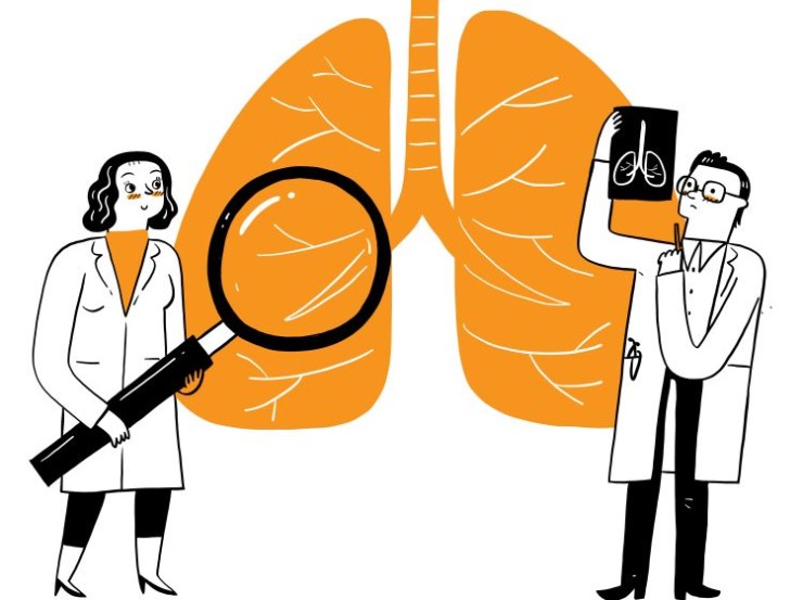
The VQ Scan is a dual-component imaging technique that combines ventilation and perfusion assessments to provide a comprehensive view of lung function.
VQ Scan or Lung Perfusion and Ventilation Scan in India with Cost
VQ Scan or Lung
Perfusion and Ventilation Scan in Detail: Decoding Pulmonary Function through
Comprehensive Imaging
In the realm of pulmonary diagnostics, the VQ (Ventilation-Perfusion) Scan, also known as the Lung Perfusion and Ventilation Scan, stands as a pivotal tool for evaluating lung function and detecting various respiratory conditions. This detailed guide aims to unravel the significance, procedure, and insights provided by VQ scans, shedding light on their applications in assessing pulmonary health.
Introduction
The VQ Scan is a dual-component imaging technique that combines ventilation and perfusion assessments to provide a comprehensive view of lung function. This non-invasive diagnostic tool is particularly valuable in identifying pulmonary embolism, assessing lung ventilation, and aiding in the diagnosis of respiratory disorders.
Understanding VQ Scan
Imaging
Ventilation Scan:
The ventilation component involves inhaling a radiolabeled gas, typically xenon or technetium, which is then imaged to assess airflow and distribution in the lungs.
Perfusion Scan:
The perfusion aspect entails injecting a radiotracer, often technetium, into the bloodstream. This substance travels to the pulmonary vasculature and is imaged to evaluate blood flow and assess lung perfusion.
Importance in Pulmonary
Imaging
The VQ Scan holds significant importance in various pulmonary evaluations:
Pulmonary Embolism Detection: Identifying
blood clots in the pulmonary arteries, a crucial diagnostic application.
Ventilation Assessment: Evaluating the
distribution of inhaled air to detect airflow abnormalities.
Perfusion Abnormalities: Assessing blood
flow to identify regions with compromised perfusion.
Chronic Lung Conditions: Aiding in the
diagnosis and monitoring of chronic lung diseases such as chronic obstructive
pulmonary disease (COPD).
Preparation for the Scan
Preparation for a VQ Scan typically involves:
No Fasting: Patients usually do not need to
fast before the scan.
No Special Diet: There are no specific dietary
restrictions.
Procedure: Comprehensive
Lung Evaluation
Ventilation Imaging: The patient inhales the radiolabeled gas, and a
gamma camera captures images to assess the distribution of inhaled air in the
lungs.
Perfusion Imaging: A radiotracer
is injected into the bloodstream, and images are taken to evaluate blood flow
in the pulmonary vasculature.
Image Fusion: Ventilation and perfusion images
are often fused to provide a comprehensive assessment of lung function.
Assessment Areas in VQ
Scan Imaging
VQ Scans are employed to assess various aspects of pulmonary health, including:
Pulmonary Embolism Detection: Identifying
areas of reduced or absent perfusion indicative of blood clots.
Ventilation Abnormalities: Detecting areas
with impaired airflow or ventilation.
Perfusion Abnormalities: Assessing blood
flow to identify regions with compromised perfusion.
Benefits of VQ Scan
Imaging
Comprehensive Evaluation: Provides a holistic
assessment of both ventilation and perfusion in the lungs.
Pulmonary Embolism Diagnosis: Crucial in
identifying and locating blood clots in the pulmonary vasculature.
Non-Invasive Nature: A non-invasive
alternative to other pulmonary diagnostic procedures.
Risks and Considerations
VQ Scans involve exposure to low levels of radiation. However, the benefits of accurate pulmonary evaluation generally outweigh the associated risks.
Clinical Applications
VQ Scans find applications in various clinical scenarios, including:
Emergency Room Settings: Rapid
evaluation of suspected pulmonary embolism.
Chronic Respiratory Conditions: Assessing lung
function in patients with chronic respiratory diseases.
Preoperative Assessment: Screening for
pulmonary complications before certain surgeries.
Expert Perspectives
Radiologists, pulmonologists, and nuclear medicine specialists collaborate to interpret VQ Scan results, providing expert insights into pulmonary function and potential abnormalities.
Technological Advancements
Ongoing advancements in imaging technology contribute to the refinement of VQ Scans, enhancing image resolution and diagnostic accuracy.
Patient Experience
VQ Scans are generally well-tolerated by patients, involving minimal discomfort. The procedure provides valuable information to healthcare providers without invasive measures.
Conclusion
In conclusion, the VQ Scan, or Lung Perfusion and Ventilation Scan, emerges as a vital tool in pulmonary diagnostics, offering a dual perspective on lung function. Its applications in detecting pulmonary embolism, assessing ventilation, and identifying perfusion abnormalities contribute to informed decision-making in respiratory care.
Frequently Asked
Questions (FAQs) related to VQ Scan or Lung Perfusion and Ventilation Scan:
1. How long does a VQ Scan typically take?
The duration of a VQ Scan can vary, but it usually takes approximately 30 minutes to an hour. This includes both the ventilation and perfusion imaging components.
2. Is there any preparation required before a VQ Scan?
Unlike some imaging procedures, VQ Scans generally require minimal preparation. Patients are usually not required to fast, and there are no specific dietary restrictions. However, it's essential to inform your healthcare provider about any pre-existing conditions and medications.
3. Are there any side effects or discomfort associated with VQ Scans?
VQ Scans are considered safe and generally well-tolerated. The most common discomfort is associated with the injection of the radiotracer, which might cause a brief sensation. Allergic reactions to the radiotracers are rare but possible. It's crucial to inform the healthcare team of any allergies beforehand.
4. Can pregnant or breastfeeding women undergo a VQ Scan?
While VQ Scans involve exposure to low levels of radiation, the risks to the fetus or breastfeeding infant are generally considered minimal. However, it's essential to inform the healthcare provider if you are pregnant or breastfeeding to assess the necessity and potential risks.
5. How often are VQ Scans recommended for monitoring chronic respiratory conditions?
The frequency of VQ Scans for monitoring chronic respiratory conditions depends on the specific medical condition and the healthcare provider's recommendations. In many cases, these scans are performed periodically to assess disease progression or treatment effectiveness.
6. Is the radiation exposure from a VQ Scan dangerous?
The radiation dosage employed in VQ Scans is deemed safe for diagnostic purposes. Typically, the advantages of acquiring precise diagnostic information are more significant than the minimal risks linked to exposure to radiation. Nevertheless, it is essential to address any concerns by having a discussion with your healthcare provider.
7. Can VQ Scans be performed on pediatric patients?
Yes, VQ Scans can be performed on pediatric patients when necessary. The radiation dose is adjusted based on the child's size, and the procedure is tailored to meet pediatric imaging standards.
8. What information does a VQ Scan provide that other lung imaging techniques may not?
VQ Scans provide a unique combination of ventilation and perfusion information in a single scan. This dual assessment allows for a comprehensive evaluation of lung function, aiding in the detection of conditions such as pulmonary embolism and ventilation abnormalities.
9. Are there alternatives to VQ Scans for assessing lung function?
While VQ Scans offer a comprehensive assessment, alternatives such as chest X-rays, CT scans, and pulmonary function tests are used for specific aspects of lung evaluation. The choice of imaging method depends on the clinical scenario and the information needed.
10. What happens after the VQ Scan?
Following the completion of the scan, patients are free to return to their regular activities. The healthcare provider will analyze the results, engage in discussions about the findings, explore their implications, and provide guidance on any required follow-up measures or treatments.
Remember, these FAQs provide general information, and specific details may vary based on individual cases and healthcare provider recommendations. Always consult with your healthcare team for personalized information regarding your medical condition and diagnostic procedures.
(0)
Login to continue


