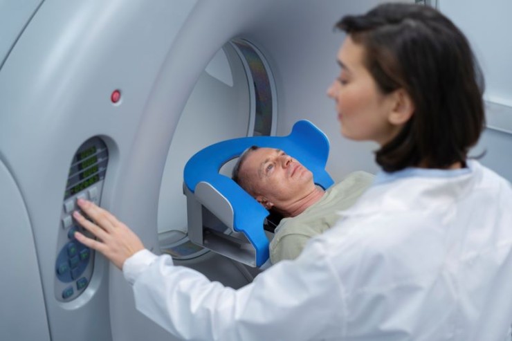
The PSMA PET-CT Scan represents a cutting-edge imaging technique designed to enhance the detection, localization, and staging of prostate cancer.
PSMA PET-CT Scan in India with Cost
Detailed Exploration of PSMA PET-CT Scan:
Transforming Prostate Cancer Imaging
In the realm of advanced medical imaging, the PSMA PET-CT
(Prostate-Specific Membrane Antigen Positron Emission Tomography-Computed
Tomography) Scan stands as a groundbreaking diagnostic tool, particularly in
the field of prostate cancer. This comprehensive guide aims to elucidate the
significance, procedure, and applications of the PSMA PET-CT Scan, providing a
detailed understanding of its role in modern healthcare.
Introduction
The PSMA PET-CT Scan represents a
cutting-edge imaging technique designed to enhance the detection, localization,
and staging of prostate cancer. By targeting the Prostate-Specific Membrane
Antigen (PSMA), this dual-imaging modality offers unprecedented precision in
assessing prostate cancer activity and spread.
Understanding PSMA PET-CT Imaging
Prostate-Specific Membrane Antigen (PSMA):
PSMA is a protein abundantly expressed in prostate cancer cells. The PSMA
PET-CT Scan utilizes a radiolabeled ligand that selectively binds to PSMA,
enabling accurate visualization of prostate cancer cells.
Positron Emission Tomography (PET):
In the PET component, the radiolabeled PSMA
ligand emits positrons, which combine with electrons in the body, producing
gamma rays. The PET scanner detects these gamma rays, mapping the distribution
of PSMA in the prostate and potentially metastatic sites.
Computed Tomography (CT):
The CT component provides detailed
anatomical images, offering structural context to the PET findings. The fusion
of PET and CT images creates a comprehensive representation.
Importance in Diagnostic Imaging
The PSMA PET-CT Scan holds paramount importance in various aspects of
prostate cancer management:
Cancer Detection and Localization: Precise imaging of prostate cancer cells, aiding in
detection and localization.
Staging and Metastasis Identification: Accurate assessment of cancer spread, especially to
distant sites, facilitating proper staging.
Treatment Planning and Response Monitoring: Assisting in treatment planning and monitoring the
response to therapies, including targeted treatments.
Preparation for PSMA PET-CT Scan
Preparation for a PSMA PET-CT Scan typically involves:
Fasting: Patients may be required to fast before the scan for optimal imaging
results.
Hydration: Adequate hydration before and after the scan helps flush the
radiotracer through the system.
Medication Adjustments: Temporary cessation or adjustment of certain
medications that may interfere with the scan.
Procedure: Illuminating Prostate Cancer Activity
Radiotracer Injection: The patient receives the radiolabeled PSMA ligand
intravenously, allowing it to circulate and bind to PSMA in prostate cancer
cells.
Uptake Period: The radiotracer accumulates in prostate cancer
cells, reaching peak concentration during a specific waiting period.
Imaging Process: The combined PET and CT scanner captures images,
creating a fused representation that overlays metabolic and anatomical details,
highlighting PSMA activity.
Assessment Areas in PSMA PET-CT Scan
PSMA PET-CT Scans are employed to assess various aspects of prostate
cancer:
Primary Tumor Imaging: Precise visualization of the primary prostate tumor.
Lymph Node Involvement: Detection and staging of cancer in nearby lymph
nodes.
Metastasis Identification: Identifying distant metastases, aiding in proper
staging.
Benefits of PSMA PET-CT Scan
Enhanced Sensitivity and Specificity: Superior sensitivity
in detecting prostate cancer cells, leading to more accurate diagnoses.
Detailed Staging: Provides detailed information about the extent of
cancer spread for comprehensive staging.
Treatment Guidance: Assists in planning targeted treatments and
monitoring treatment responses.
Risks and Considerations
The PSMA PET-CT Scan involves exposure to low levels of radiation. However,
the benefits of precise prostate cancer assessment generally outweigh the
associated risks.
Clinical Applications
PSMA PET-CT Scans find applications in various clinical scenarios,
including:
Prostate Cancer Diagnosis: Enhancing accuracy in detecting and localizing
prostate cancer.
Staging and Monitoring: Facilitating proper staging and monitoring treatment
responses.
Expert
Perspectives
Radiologists, urologists, and oncologists
collaborate to interpret PSMA PET-CT Scan results, providing expert insights
into prostate cancer activity.
Technological Advancements
Continual advancements in imaging technology
contribute to the refinement of PSMA PET-CT Scans, enhancing image resolution
and diagnostic capabilities.
Patient Experience
While the PSMA PET-CT Scan involves exposure
to radiation, it is generally well-tolerated by patients. The procedure
provides valuable information to healthcare providers without invasive
measures.
Conclusion
In conclusion, the PSMA PET-CT Scan
represents a significant advancement in prostate cancer imaging, offering
unparalleled precision in detecting and assessing the activity of prostate
cancer cells. Its applications in cancer detection, staging, and treatment
monitoring contribute to advanced and personalized healthcare for individuals
with prostate cancer.
Frequently Asked
Questions (FAQs) About PSMA PET-CT Scan
1. What is PSMA, and why is it significant in prostate cancer imaging?
PSMA, or Prostate-Specific Membrane Antigen, is a protein highly expressed
in prostate cancer cells. PSMA PET-CT leverages this specificity to precisely
locate and visualize prostate cancer cells, enhancing diagnostic accuracy.
2. How does a PSMA PET-CT Scan differ from
traditional imaging methods for prostate cancer?
PSMA PET-CT offers superior sensitivity and
specificity in detecting prostate cancer cells compared to traditional imaging
methods. It provides detailed metabolic and anatomical information, aiding in
precise diagnosis and staging.
3. Is the PSMA PET-CT Scan painful or
invasive?
No, the PSMA PET-CT Scan is a non-invasive
procedure. Patients may experience mild discomfort during the intravenous
injection of the radiotracer, but the scan itself is generally well-tolerated.
4. What preparations are required before
undergoing a PSMA PET-CT Scan?
Preparations may include fasting, staying
hydrated, and temporary cessation or adjustment of specific medications. Your
healthcare provider will provide detailed instructions based on your individual
case.
5. How long does the PSMA PET-CT Scan
procedure take?
The entire procedure typically takes a few
hours, including the waiting period for the radiotracer to accumulate. However,
scan durations may vary, and your healthcare team will provide specific
details.
6. Are there any risks associated with
radiation exposure during a PSMA PET-CT Scan?
While the scan involves exposure to low levels
of radiation, the benefits of precise prostate cancer assessment generally
outweigh the associated risks. The radiation dose is carefully controlled to
minimize potential harm.
7. Can the PSMA PET-CT Scan detect cancer
spread to other parts of the body?
Yes, one of the key advantages of the PSMA
PET-CT Scan is its ability to detect metastases or cancer spread to distant
sites. This information is crucial for accurate staging and treatment planning.
8. How often should a patient undergo a PSMA
PET-CT Scan during prostate cancer treatment?
The frequency of PSMA PET-CT scans depends
on the individual's treatment plan and the need for ongoing monitoring. Your
healthcare team will determine the most suitable schedule based on your
specific case.
9. Are there any specific patient groups for
whom PSMA PET-CT is particularly beneficial?
PSMA PET-CT is especially beneficial for
individuals with suspected or confirmed prostate cancer, especially when
traditional imaging methods may present challenges or uncertainties in
diagnosis, staging, or treatment monitoring.
10. Can the PSMA PET-CT Scan influence
treatment decisions for prostate cancer?
Yes, the PSMA PET-CT Scan significantly
influences treatment decisions for prostate cancer. By providing detailed
information on the location and extent of cancer cells, it helps healthcare
professionals plan targeted treatments and assess responses, contributing to
more personalized and effective care.
These FAQs offer insights into the PSMA PET-CT
Scan, addressing common queries patients may have about this advanced
diagnostic imaging technique for prostate cancer. Always consult with your
healthcare provider for personalized information based on your specific medical
situation.
(0)
Login to continue


