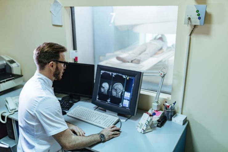
CT Brain and Venography with Contrast amalgamate neuroimaging and vascular assessment, providing detailed insights into both brain structures and venous circulation.
CT Brain and Venography Scan with Contrast in India with Cost
CT Brain and Venography
with Contrast: Navigating Neurovascular Precision
Introduction
Delving into Neurovascular Insights with CT Brain and Venography with Contrast
CT Brain and Venography with Contrast amalgamate neuroimaging and vascular assessment, providing detailed insights into both brain structures and venous circulation. This comprehensive guide aims to elucidate the significance, procedure, and applications of CT Brain and Venography with Contrast, offering a thorough understanding of its role in neurovascular diagnostics.
Understanding CT Brain and Venography with Contrast
What is CT Brain and Venography with Contrast?
CT Brain and Venography with Contrast is an advanced imaging technique that utilizes computed tomography to capture high-resolution images of both the brain and venous structures, enhancing visibility through the administration of contrast agents. This specialized scan allows healthcare professionals to assess neurovascular structures, detect abnormalities, and make precise diagnoses.
The Significance of CT
Brain and Venography with Contrast
Integrated Neurovascular Imaging
CT Brain and Venography with Contrast play a pivotal role in achieving integrated neurovascular imaging, offering detailed views of the brain's structures and venous circulation. This enables the identification of conditions such as vascular malformations, thrombosis, or tumors affecting both neurological and vascular health.
Comprehensive Vascular Assessment
With the use of contrast agents, the scan facilitates a comprehensive assessment of venous structures, allowing healthcare professionals to identify abnormalities affecting blood flow, clotting, or venous drainage.
The Procedure: A Step-by-Step Guide
Patient Preparation
Patients undergoing CT Brain and Venography with Contrast typically require minimal preparation. It's crucial to communicate any allergies, kidney issues, or relevant medical history to the healthcare team.
Contrast Administration and Image Acquisition
Contrast dye is administered intravenously, and the CT scanner captures multiple high-resolution images, providing detailed cross-sectional views of both the brain and venous structures with enhanced contrast visibility.
Applications of CT Brain and Venography with Contrast
Detecting Neurovascular Abnormalities
CT Brain and Venography with Contrast are highly effective in detecting neurovascular abnormalities, offering precise images that assist in determining the location, extent, and characteristics of conditions affecting both the brain and venous circulation.
Identifying Venous Thrombosis and Tumors
The detailed imaging capabilities of this scan make it an invaluable tool for identifying venous thrombosis, tumors, or other abnormalities affecting both neurological and vascular health, contributing to accurate diagnoses.
Advantages and Considerations
Advantages of CT Brain and Venography with Contrast
Integrated Neurovascular Visualization
CT Brain and Venography with Contrast provide integrated visualization of neurovascular structures, ensuring accurate assessment and aiding in the identification of various conditions affecting both systems.
Comprehensive Vascular Assessment
The procedure offers a comprehensive assessment of venous structures, allowing for a thorough understanding of neurovascular health.
Considerations and Precautions
Contrast-Related Considerations
While generally safe, the medical team considers any potential allergies or kidney issues that might affect contrast utilization.
Radiation Exposure
As with any CT scan, consideration is given to radiation exposure. The healthcare team carefully evaluates the benefits against the risks for each patient, prioritizing patient safety.
Conclusion
In conclusion, CT Brain and Venography with Contrast emerge as crucial diagnostic tools, providing detailed and high-resolution images for the precise assessment of both neurological and vascular conditions. From venous thrombosis to neurovascular tumors, this integrated imaging technique significantly contributes to enhancing overall health diagnostics.
FAQs: Clarifying CT Brain and Venography with Contrast
1. Is CT Brain and Venography with Contrast a painful procedure?
No, the procedure is generally well-tolerated. Patients may experience a sensation of warmth during contrast injection.
2. How long does a CT Brain and Venography with Contrast take?
The procedure typically takes around 30 to 60 minutes, depending on the patient's condition.
3. Are there any dietary restrictions before a CT Brain and Venography with Contrast?
Generally, there are no specific dietary restrictions for this scan. However, patients are advised to follow any instructions provided by the healthcare team.
4. Can pregnant women undergo CT Brain and Venography with Contrast?
While generally avoided during pregnancy, in specific cases, the benefits and risks are carefully evaluated to make an informed decision.
5. How often is CT Brain and Venography with Contrast recommended for follow-up examinations?
Follow-up scans are scheduled based on the individual's medical history and the presence of specific neurovascular conditions.
6. Is there any special preparation required before a CT Brain and Venography with Contrast?
Generally, minimal preparation is needed. However, inform your healthcare team about any allergies, medications, or existing health conditions.
7. Are there any risks associated with the contrast dye used in the procedure?
While contrast dye is generally safe, some individuals may experience mild allergic reactions. Your healthcare team will assess potential risks based on your medical history.
8. Can individuals with kidney issues undergo CT Brain and Venography with Contrast?
Special consideration is given to individuals with kidney problems. Your healthcare team will assess the risks and benefits, and alternative imaging methods may be considered.
9. Is CT Brain and Venography with Contrast suitable for children?
The procedure can be performed on children, but the decision depends on the specific case. Pediatric imaging protocols may be adjusted to ensure safety and accuracy.
10. How soon can I resume normal activities after CT Brain and
Venography with Contrast?
While many patients can promptly return to their regular activities post-procedure, it is recommended to have a companion accompany them home, particularly if sedation is administered.
(0)
Login to continue


