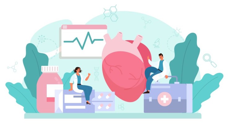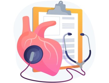
A 2D Echo, or two-dimensional echocardiogram, is a non-invasive imaging test that uses sound waves to create detailed images of the heart's structure and function.
2D Echo Test with Cost
Unveiling the Wonders of 2D Echo Test: A
Comprehensive Guide
Introduction to 2D Echo Test
A 2D Echo, or two-dimensional echocardiogram, is a non-invasive imaging test that uses sound waves to create detailed images of the heart's structure and function. This article provides a comprehensive guide to understanding the intricacies of this cardiac diagnostic procedure.
Significance of 2D Echo
in Cardiac Evaluation
2D Echo plays a crucial role in cardiac evaluation, offering valuable insights into the heart's chambers, valves, and overall function. It aids in diagnosing various cardiovascular conditions, providing essential information for treatment planning.
Preparation for the Procedure
Patients undergoing a 2D Echo may need to follow specific preparation guidelines. This typically includes fasting for a few hours before the test and avoiding certain medications that could affect heart rate or blood vessels.
Procedure Overview
During the 2D Echo procedure, a transducer is placed on the chest to transmit sound waves, which bounce back to create detailed images of the heart on a monitor. This real-time imaging allows healthcare professionals to assess the heart's structure and function.
Components of 2D Echo Test
The 2D Echo test comprises several components, including M-mode imaging, Doppler imaging, and color Doppler imaging. These components provide different perspectives and additional information about the heart's performance.
Advantages of 2D Echo
over Traditional Imaging
2D Echo offers distinct advantages over traditional imaging methods. It is non-invasive, radiation-free, and provides real-time imaging, allowing for a comprehensive evaluation of the heart without exposing patients to ionizing radiation.
Common Indications for 2D Echo
Healthcare providers may recommend a 2D Echo for various indications, including assessing heart function, detecting structural abnormalities, evaluating valve conditions, and monitoring the progression of cardiac diseases.
Interpretation of Results
Interpreting 2D Echo results requires expertise in cardiac imaging. Radiologists and cardiologists analyze the images to identify abnormalities, measure chamber dimensions, assess valve function, and determine overall cardiac health.
Risks and Safety Measures
2D Echo is considered a safe procedure with minimal risks. However, patients with certain medical conditions or allergies to ultrasound gel should inform their
healthcare providers. Stringent safety protocols are implemented to guarantee the well-being of the patient.
Comparisons with Other Cardiac Imaging Techniques
Comparing 2D Echo with other cardiac imaging techniques, such as CT scans or MRI, highlights the unique advantages of each method. Factors like accessibility, cost, and the specific diagnostic goals influence the choice of imaging modality.
Innovations and Advancements in 2D Echo
Technological advancements continue to enhance the capabilities of 2D Echo. Innovations, such as three-dimensional echocardiography and strain imaging, contribute to increased diagnostic accuracy and a deeper understanding of cardiac conditions.
Patient Experience and
Comfort
The non-invasive nature of 2D Echo contributes to a comfortable patient experience. The procedure is painless, and patients can typically resume their normal
activities immediately after the test.
Follow-up Care and Recommendations
After undergoing a 2D Echo, patients receive detailed reports from their healthcare providers. Follow-up care may involve additional tests or consultations based on the findings, ensuring comprehensive and personalized medical attention.
Case Studies and Success Stories
Real-life case studies and success stories highlight the effectiveness of 2D Echo in diagnosing and monitoring cardiac conditions. These narratives provide insights into the impact of timely imaging on patient outcomes.
Expert Insights and Recommendations
Leading experts in cardiac imaging share their insights and recommendations on the evolving landscape of 2D Echo. Their perspectives on best practices, ongoing research, and future developments contribute to the continuous improvement of this diagnostic modality.
Conclusion
In conclusion, the 2D Echo test stands as a valuable diagnostic tool in the realm of cardiac evaluation. Its non-invasive nature, coupled with technological advancements, makes it an indispensable method for assessing and monitoring various cardiovascular conditions.
FAQs (Frequently Asked Questions) about 2D Echo Test
Is the 2D Echo test painful?
No, the 2D Echo test is a non-invasive procedure and is generally painless. The transducer is placed on the chest, and patients may feel slight pressure but no discomfort.
How long does a 2D Echo test take?
The duration of a 2D Echo test varies but typically takes around 30 to 60 minutes, depending on the complexity of the examination.
Can pregnant women undergo a 2D Echo test?
Yes, 2D Echo is considered safe for pregnant women when medically necessary. However, the healthcare provider will weigh the benefits against potential risks.
Are there any side effects of a 2D Echo test?
Generally, there are no significant side effects. The procedure uses sound waves and does not involve ionizing radiation, minimizing risks.
Is a 2D Echo capable of detecting various heart conditions?
While 2D Echo is effective in diagnosing many heart conditions, some complex or specific conditions may require additional imaging methods for a comprehensive evaluation.
Is eating or drinking allowed before a 2D Echo test?
It's generally recommended to fast for a few hours before a 2D Echo test to minimize interference from digestive activities. Your healthcare provider will provide specific instructions based on your individual case.
How often should one undergo a routine 2D Echo screening?
The frequency of routine 2D Echo screenings depends on individual health factors and risk assessments. Your healthcare provider will determine an appropriate screening schedule based on your medical history and cardiac health.
Are there any age restrictions for a 2D Echo test?
2D Echo tests can be performed on individuals of all ages, from infants to the elderly. The procedure is safe and widely used across age groups for assessing various cardiac conditions.
Can 2D Echo detect coronary artery disease?
While 2D Echo provides valuable information about heart structures and function, it may not be the primary tool for diagnosing coronary artery disease. Other imaging modalities like CT angiography or stress tests may be recommended for assessing coronary arteries.
Is there any discomfort associated with the application of the transducer during a 2D Echo?
The application of the transducer is painless, and patients typically experience minimal discomfort. The gel used to enhance the sound wave transmission may feel cool on the skin.
(0)
Login to continue


